. 2010 Dec;16(12):1400-6.
doi: 10.1038/nm.2252. Epub 2010 Nov 21.

Conversion of vascular endothelial cells into multipotent stem-like cells

Affiliations

Conversion of vascular endothelial cells into multipotent stem-like cells

Damian Medici et al. Nat Med. 2010 Dec.

Erratum in

  • Nat Med. 2011 Apr;17(4):514

Abstract

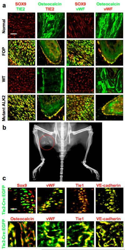
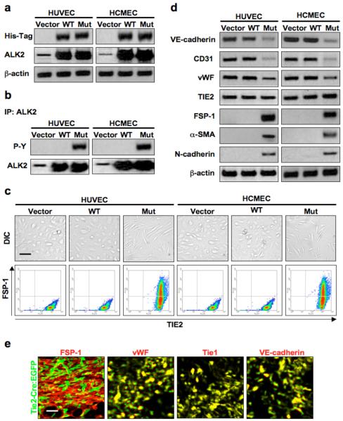
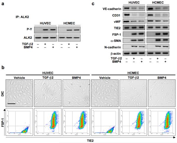
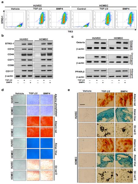
Mesenchymal stem cells can give rise to several cell types, but varying results depending on isolation methods and tissue source have led to controversies about their usefulness in clinical medicine. Here we show that vascular endothelial cells can transform into multipotent stem-like cells by an activin-like kinase-2 (ALK2) receptor-dependent mechanism. In lesions from individuals with fibrodysplasia ossificans progressiva (FOP), a disease in which heterotopic ossification occurs as a result of activating ALK2 mutations, or from transgenic mice expressing constitutively active ALK2, chondrocytes and osteoblasts expressed endothelial markers. Lineage tracing of heterotopic ossification in mice using a Tie2-Cre construct also suggested an endothelial origin of these cell types. Expression of constitutively active ALK2 in endothelial cells caused endothelial-to-mesenchymal transition and acquisition of a stem cell-like phenotype. Similar results were obtained by treatment of untransfected endothelial cells with the ligands transforming growth factor-β2 (TGF-β2) or bone morphogenetic protein-4 (BMP4) in an ALK2-dependent manner. These stem-like cells could be triggered to differentiate into osteoblasts, chondrocytes or adipocytes. We suggest that conversion of endothelial cells to stem-like cells may provide a new approach to tissue engineering.


Figures

Figure 1
Figure 1
Endothelial ceΠ differentiation in heterotopic ossification. (a) Immunohistochemistry of chondrogenic (first and third columns from left) and osteogenic (second and forth columns from left) lesions from Fibrodysplasia Ossificans Progressiva (FOP) patients with activating ALK2 mutations and from Cre-dependent constitutively active ALK2 (caALK2) transgenic mice. Chondrogenic lesions show co-expression of the endothelial markers TIE2 and vWF with the chondrocyte marker SOX9. Osteogenic lesions show co-expression of TIE2 and vWF with the osteoblast marker osteocalcin. Normal cartilage and bone from human hip joint (top row) or wild-type (WT) mouse knee joint (third row) show no evidence of TIE2- or vWF-positive chondrocytes or osteoblasts. Scale bar, 40 μm. (b) X-ray image of heterotopic ossification (red circle) in a Cre-induced caALK2 transgenic mouse. (c) Immunohistochemistry of BMP4-induced heterotopic cartilage and bone in Tie2-Cre reporter mice showing EGFP positive chondrocytes (top row) and osteoblasts (bottom row). These EGFP positive cells show expression of endothelial markers vWF, Tie1, and VE-cadherin. Scale bar, 20 μm.
Figure 2
Figure 2
Constitutively active ALK2 promotes endothelial-mesenchymal transition. (a) Immunoblotting showing positive expression of the His-Tag on wild-type (WT) and mutant (Mut) ALK2 in infected endothelial cells, as well as increased expression of total ALK2. β-actin was used as an internal control. (b) Immunoprecipitation demonstrating constitutive tyrosine phosphorylation (P-Y) of the mutant ALK2 receptor. (c) DIC imaging (top row) showing a change in cell morphology in endothelial cells expressing mutant ALK2. Flow cytometry analysis (bottom row) demonstrating co-expression of TIE2 and FSP-1 in cells containing the mutant ALK2 construct. Scale bar, 10 μm. (d) Immunoblotting showing deceased expression of the endothelial markers VE-cadherin, CD31, and vWF and increased expression of the mesenchymal markers FSP-1, α-SMA, and N-cadherin in endothelial cells expressing mutant ALK2 but not in cells infected with vector or wild-type ALK2 adenoviral constructs. TIE2 levels remained constant. β-actin was used as an internal control. (e) Immunohistochemistry of early stage BMP4-induced lesions of heterotopic ossification in Tie2-Cre reporter mice showing EGFP positive mesenchymal cells. Most of these EGFP positive cells also show expression of the endothelial markers vWF, Tie1, and VE-cadherin. Scale bar, 20 μm.
Figure 3
Figure 3
Formation of endothelial derived multipotent stem-like cells induced by constitutively active ALK2. (a) Flow cytometry analysis showing co-expression of TIE2 and STRO-1 in endothelial cells expressing mutant ALK2. (b) Immunoblotting showing expression of the mesenchymal stem cell markers STRO-1, CD10, CD44, CD71, CD90, and CD117 in endothelial cells expressing mutant ALK2. Human bone marrow derived mesenchymal stem cells (MSC) express these markers, but human corneal fibroblasts (HCF) do not. β-actin was used as an internal control. (c) Immunoblotting showing increased expression of osteoblast (osterix), chondrocyte (SOX9), or adipocyte (PPARγ2) markers in cells treated with mutant ALK2 for 48 h followed by exposure to osteogenic, chondrogenic, or adipogenic culture medium. (d) Positive staining of osteoblast (alkaline phosphatase and alizarin red), chondrocyte (alcian blue), or adipocyte (oil red O) products in endothelial cell cultures treated with mutant ALK2, but not with vector or wild-type ALK2, for 48 h followed by growth in osteogenic, chondrogenic, or adipogenic culture medium, respectively. Scale bar, 100 μm. (e) Positive staining of osteoblast (alizarin red), chondrocyte (alcian blue), or adipocyte (oil red O) products in polylactic acid scaffolds containing endothelial cells transformed by mutant ALK2 implanted into nude mice, followed by local injection of osteogenic, chondrogenic, or adipogenic medium every 72 h for 6 weeks. Scale bar, 100 μm.
Figure 4
Figure 4
TGF-β2 and BMP4 activate ALK2 and induce endothelial-mesenchymal transition. (a) Immunoblotting of immunoprecipitates confirming phosphorylation of ALK2 by 15 min of TGF-β2 or BMP4 stimulation. (b) DIC imaging, immunocytochemistry and flow cytometry showing a change in cell morphology and co-expression of TIE2 and FSP-1 in endothelial cells treated with TGF-β2 or BMP4 for 48 h. Scale bar, 20 μm. (c) Immunoblotting confirming EndMT with decreased expression of VE-cadherin, CD31, and vWF and increased expression of FSP-1, α-SMA, and N-cadherin in cells treated with TGF-β2 or BMP4. TIE2 levels remained constant.
Figure 5
Figure 5
Endothelial cells transformed by treatment with TGF-β2 or BMP4 express mesenchymal stem cell markers and exhibit multipotency. (a) Flow cytometry showing co-expression of TIE2 and STRO-1 in endothelial cells treated with TGF-β2 or BMP4 for 48 h. (b) Immunoblotting confirming increased protein expression of mesenchymal stem cell markers STRO-1, CD10, CD44, CD71, CD90, and CD117 in cells treated with TGF-β2 or BMP4. β-actin was used as an internal control. (c) Immunoblotting showing increased expression of osteoblast (osterix), chondrocyte (SOX9), or adipocyte (PPARγ2) markers in cells treated with TGF-β2 or BMP4 for 48 h followed by exposure to osteogenic, chondrogenic, or adipogenic culture medium, respectively. (d) Positive staining of osteoblast (alkaline phosphatase and alizarin red), chondrocyte (alcian blue), or adipocyte (oil red O) products in endothelial cell cultures treated with TGF-β2 or BMP4 for 48 h followed by growth in osteogenic, chondrogenic, or adipogenic culture medium. Scale bar, 100 μm. (e) Positive staining of osteoblast (alizarin red), chondrocyte (alcian blue), or adipocyte (oil red O) products in polylactic acid scaffolds containing endothelial cells transformed by TGF-β2 or BMP4 subcutaneously implanted into nude mice, followed by local injection of osteogenic, chondrogenic, or adipogenic medium every 72 h for 6 weeks. Scale bar, 100 μm.
Figure 6
Figure 6
ALK2 is necessary for EndMT. (a) Immunoblotting confirming knockdown of ALK2 expression by ALK2 siRNA in HUVEC and HCMEC cultures. (b) Immunoblotting showing increased expression of FSP-1 and STRO-1 in TGF-β2 or BMP4 treated endothelial cells transfected with negative control siRNA, but inhibition of this expression in cells transfected with ALK2 siRNA. β-actin was used as an internal control. (c) Flow cytometry analysis confirming increased numbers of endothelial cells expressing FSP-1 when treated with TGF-β2 or BMP4, and inhibition of such expression in cells treated with ALK2 siRNA. (d) Positive staining of osteoblast (alkaline phosphatase [AP] and alizarin red [AR]), chondrocyte (alcian blue [AB]), or adipocyte (oil red O [OR]) products in cultures transfected with negative control siRNA and treated with TGF-β2 or BMP4 for 48 h, followed by growth in osteogenic, chondrogenic, or adipogenic culture medium. In contrast, expression of ALK2 siRNA prevented the differentiation of these cells. Scale bar, 100 μm.

Comment in

References

    1. Potenta S, Zeisberg E, Kalluri R. The role of endothelial-to-mesenchymal transition in cancer progression. Br. J. Cancer. 2008;99:1375–1379. - PMC - PubMed
    1. Akhurst RJ, Derynck R. TGF-beta signaling in cancer - a double-edged sword. Trends Cell Biol. 2001;11:S44–S51. - PubMed
    1. Thiery JP. Epithelial-mesenchymal transitions in tumor progression. Nat. Rev. Cancer. 2002;2:442–454. - PubMed
    1. Thiery JP. Epithelial-mesenchymal transitions in development and pathologies. Curr. Opin. Cell Biol. 2003;15:740–746. - PubMed
    1. Hay ED. The mesenchymal cell, its role in the embryo, and the remarkable signaling mechanisms that create it. Dev. Dyn. 2005;233:706–720. - PubMed

Publication types

MeSH terms