. 2010 Dec;31(12):1569-75.
doi: 10.1038/aps.2010.176. Epub 2010 Nov 22.

Tanshinone IIA attenuates angiotensin II-induced apoptosis via Akt pathway in neonatal rat cardiomyocytes

Affiliations

Tanshinone IIA attenuates angiotensin II-induced apoptosis via Akt pathway in neonatal rat cardiomyocytes

Hong-jye HONG et al. Acta Pharmacol Sin. 2010 Dec.

Abstract

Aim: to examine the effects of tanshinone IIA, the main effective component of Salvia miltiorrhiza (known as 'Danshen' in traditional Chinese medicine) on angiotensin II (Ang II)-mediated cardiomyocyte apoptosis.

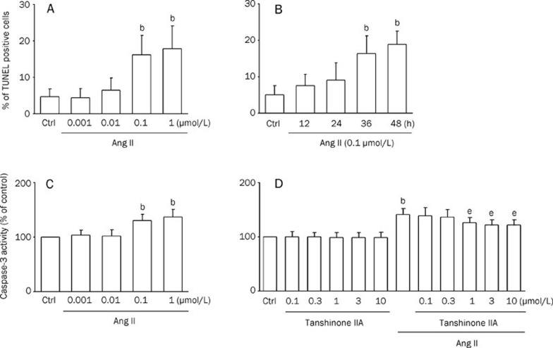
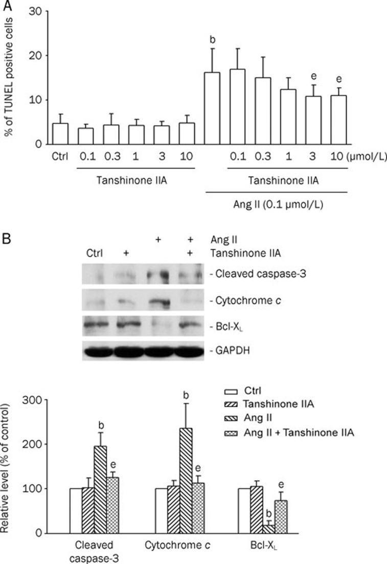
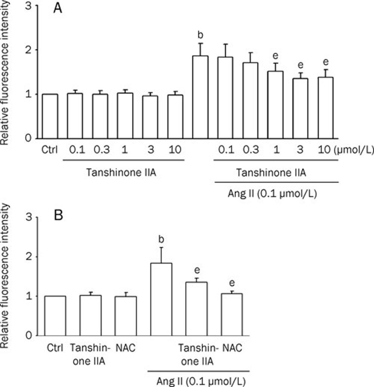
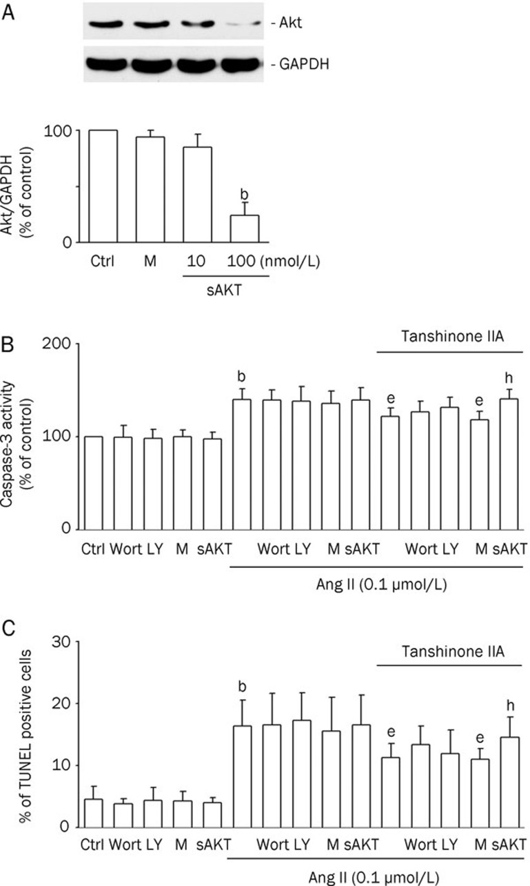
Methods: rat neonatal cardiomyocytes were primarily cultured with Ang II or Ang II plus tanshinone IIA. Myocyte apoptosis was evaluated by caspase-3 activity and DNA strand break level with TdT-mediated dUTP nick-end labeling (TUNEL) staining. Western blot analysis was employed to determine the related protein expression and flow cytometry assay was used to determine the TUNEL positive cells and the intracellular reactive oxygen species (ROS) production. SiRNA targeted to Akt was used.

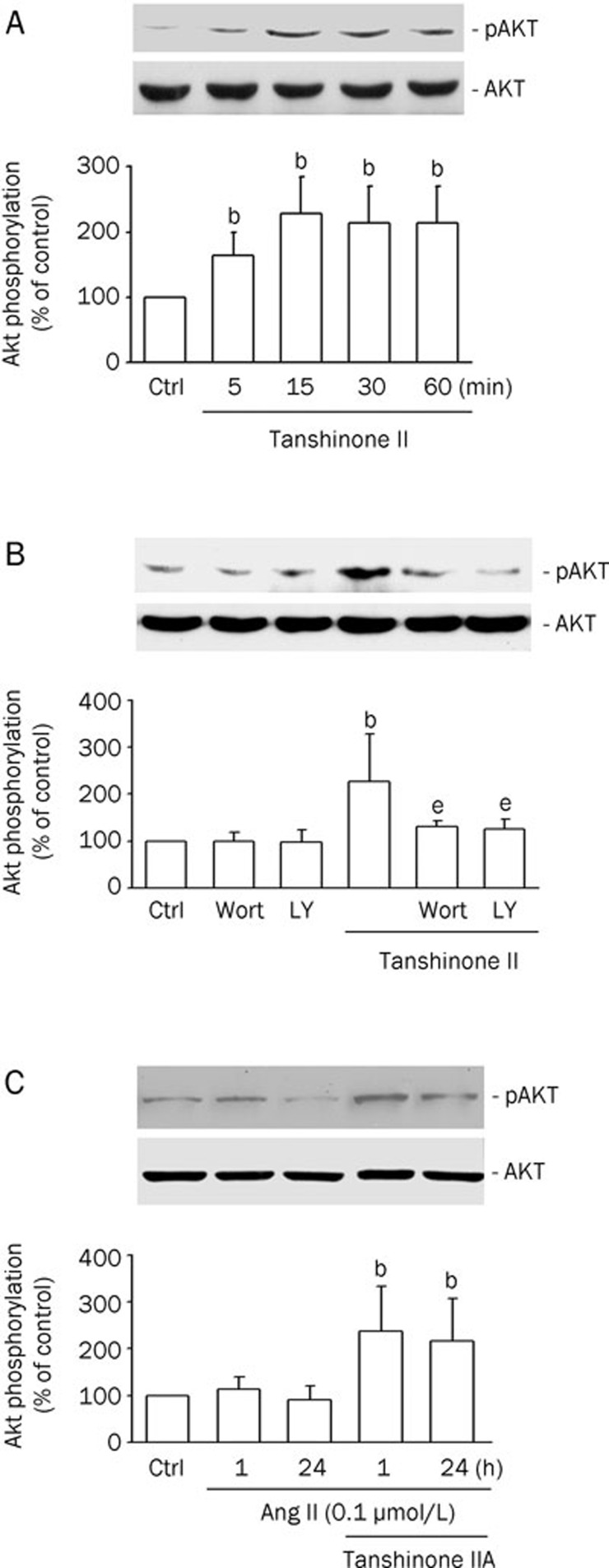
Results: ang II (0.1 micromol/L) remarkably increased caspase-3 activity, TUNEL positive cells, and cleaved caspase-3 and cytochrome c expression, but reduced Bcl-X(L) expression. These effects were effectively antagonized by pretreatment with tanshione IIA (1-3 micromol/L). Tanshinone IIA had no effect on basal ROS level, while attenuated the ROS production by Ang II. Interestingly, tanshione IIA significantly increased the phosphorylated Akt level, which was countered by the PI3K antagonist wortmannin or LY294002. Knockdown of Akt with Akt siRNA significantly reduced Akt protein levels and tanshinone IIA protective effect.

Conclusion: tanshinone IIA prevents Ang II-induced apoptosis, thereby suggesting that tanshinone IIA may be used for the prevention of the cardiac remodeling process.


Figures

Figure 1
Figure 1
Caspase-3 activity in cardiomyocytes is inhibited by tanshinone IIA. Results were shown as mean±SEM. bP<0.05 vs control (Ctrl); eP<0.05 vs Ang II. (A) Bar graph showing the percentage of cardiomyocytes undergoing apoptosis in the presence of Ang II (0.001, 0.01, 0.1, and 1 μmol/L) for 48 h. TdT-mediated dUTP nick-end labeling (TUNEL) analysis was performed as described in Materials and methods. (n=5) (B) Bar graph showing the percentage of cardiomyocytes undergoing apoptosis in the presence of Ang II (0.1 μmol/L) at different incubation times (12, 24, 36, and 48 h). (n=4) (C) Effects of angiotensin II (Ang II) (0.001, 0.01, 0.1, and 1 μmol/L) on caspase-3 activity in cardiomyocytes. Caspase-3 activity was measured in lysates prepared from cardiomyocytes. (n=4) (D) Cardiomyocytes pretreated with tanshinone IIA (0.1, 0.3, 1, 3, and 10 μmol/L; for 30 min) in the absence or presence of 0.1 μmol/L Ang II for 48 h. Bars indicate the intensity of R110 from 6 independent experiments, each in triplicate measurements. (n=8)
Figure 2
Figure 2
Tanshinone IIA protected cardiomyocytes from angiotensin II (Ang II)-induced apoptosis. (A) Flow cytometric analysis of TdT-mediated dUTP nick-end labeling (TUNEL)-stained cells. Cardiomyocytes pretreated with tanshinone IIA (0.1, 0.3, 1, 3, and 10 μmol/L; for 30 min) in the absence or presence of 0.1 μmol/L Ang II for 48 h. Percentages of apoptotic cardiomyocytes in the different groups. Results were shown as mean±SEM. (n=6). bP<0.05 vs control (Ctrl); eP<0.05 vs Ang II. (B) Effects of tanshinone IIA on apoptotic markers (cleaved caspase-3, released cytochrome c, and BcL-xL) in Ang II-treated cardiomyocytes. The cells were pretreated with tanshinone IIA (3 μmol/L) for 30 min, and then treated with 0.1 μmol/L Ang II for 12 h. Upper panels: Western blotting was carried out with the specific antibody against cleaved caspase-3, cytochrome c, and Bcl-xL. Glyceraldehyde-3-phosphate dehydrogenase (GAPDH) was used as loading control. Lower panels: The quantitative results from the Western blots. Results were shown as mean±SEM. (n=4). bP<0.05 vs control (Ctrl); eP<0.05 vs Ang II.
Figure 3
Figure 3
Effects of tanshinone IIA on angiotensin II (Ang II)-induced ROS generation in cardiomyocytes. Relative fluorescence intensity in rat cardiomyocytes was quantified by flow cytometry using dichlorofluorescin diacetate (DCFH-DA). The fluorescence intensities in untreated control cells are expressed as 100%. Data were presented as relative intensity of the experimental groups compared to untreated control cells. Results were shown as mean±SEM. (n=6). bP<0.05 vs control (Ctrl); eP<0.05 vs Ang II. (A) Column bar graph of mean cell fluorescence for dichlorofluorescein (DCF) evaluated for cardiomyocytes pretreated with tanshinone IIA (0.1, 0.3, 1, 3, and 10 μmol/L; for 30 min) and thereafter in the absence or presence of 0.1 μmol/L Ang II for 1 h. (B) Cells were incubated with tanshinone IIA (3 μmol/L) or N-acetylcysteine (NAC) (5 mmol/L) and thereafter in the absence or presence of 0.1 μmol/L Ang II for 1 h.
Figure 4
Figure 4
Tanshinone IIA induces Akt phosphorylation via PI3K. (A) Neonatal cardiomyocytes were incubated with 3 μmol/L tanshinone IIA for the indicated times. (B) Neonatal cardiomyocytes were incubated with wortmannin (Wort; 100 nmol/L) or LY294002 (LY; 10 nmol/L) for 30 min followed by incubation with 3 μmol/L tanshinone IIA for 15 min. (C) Neonatal cardiomyocytes were incubated with tanshinone IIA (3 μmol/L) for 30 min prior to the addition of Ang II (0.1 μmol/L) for 1 or 24 h. Western blot analyses were performed using site- and phospho-specific Akt antibodies against Ser473 (p-Akt, upper blot) or total Akt (lower blot). The results were shown as means±SEM (n=4), expressed as percentage changes in phosphorylation over that in control cells. bP<0.05 vs control (Ctrl). eP<0.05 vs tanshinone IIA.
Figure 5
Figure 5
Blockage of the Akt pathway attenuated the inhibitory effect of tanshinone IIA on angiotensin II (Ang II)-induced apoptosis. Notes: Ctrl, untransfected control; M, mock control; sAkt, Akt siRNA transfection. bP<0.05 vs the mock control. eP<0.05 vs the Ang II treatment. hP<0.05 vs the tanshinone IIA and Ang II treatment. (A) The effect of Akt siRNA transfection on Akt protein levels in cardiomyocytes. The cells were transfected with Akt siRNA (sAkt; 10, or 100 nmol/L) to get Akt knockdown cells. Control siRNA was also applied as mock controls (M). Western blotting was carried out with the specific antibody against Akt. GAPDH was used as a loading control. Results were shown as mean±SEM (n=3). (B) The effect of wortmannin (Wort), LY294002 (LY), and Akt siRNA on tanshinone IIA-decreased Ang II-induced caspase-3 activity in cardiomyocytes. Transfected cells were pretreated with or without tanshinone IIA (3 μmol/L) for 30 min, and then treated with Ang II (0.1 μmol/L) for 12 h. Results were shown as mean±SEM (n=6). (C) The effect of Wort, LY, and Akt siRNA on tanshinone IIA-decreased Ang II-induced apoptosis in cardiomyocytes. Transfected cells were pretreated with or without tanshinone IIA (3 μmol/L) for 30 min, and then treated with Ang II (0.1 μmol/L) for 48 h. Results were shown as mean±SEM (n=6).

Similar articles

Cited by

References

    1. Masri C, Chandrashekhar Y. Apoptosis: a potentially reversible, meta-stable state of the heart. Heart Fail Rev. 2008;13:175–9. - PubMed
    1. Satoh M, Matter CM, Ogita H, Takeshita K, Wang CY, Dorn GW, 2nd, et al. Inhibition of apoptosis-regulated signaling kinase-1 and prevention of congestive heart failure by estrogen. Circulation. 2007;115:3197–204. - PMC - PubMed
    1. Sorescu D, Griendling KK. Reactive oxygen species, mitochondria, and NAD(P)H oxidases in the development and progression of heart failure. Congest Heart Fail. 2002;8:132–40. - PubMed
    1. Paul M, Poyan Mehr A, Kreutz R. Physiology of local renin-angiotensin systems. Physiol Rev. 2006;86:747–803. - PubMed
    1. Schroder D, Heger J, Piper HM, Euler G. Angiotensin II stimulates apoptosis via TGF-beta1 signaling in ventricular cardiomyocytes of rat. J Mol Med. 2006;84:975–83. - PubMed

Publication types

MeSH terms