

Temporal spatial expression and function of non-muscle myosin II isoforms IIA and IIB in scar remodeling
- PMID: 21102503
- PMCID: PMC3407540
- DOI: 10.1038/labinvest.2010.181
Temporal spatial expression and function of non-muscle myosin II isoforms IIA and IIB in scar remodeling
Abstract
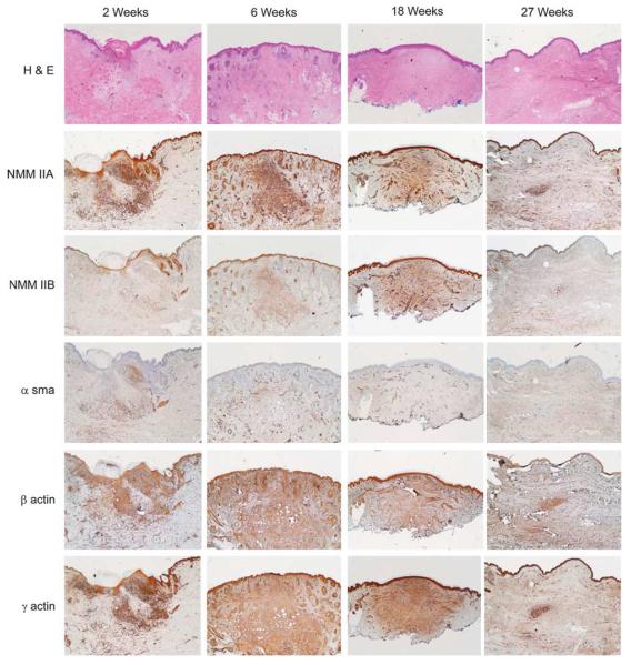
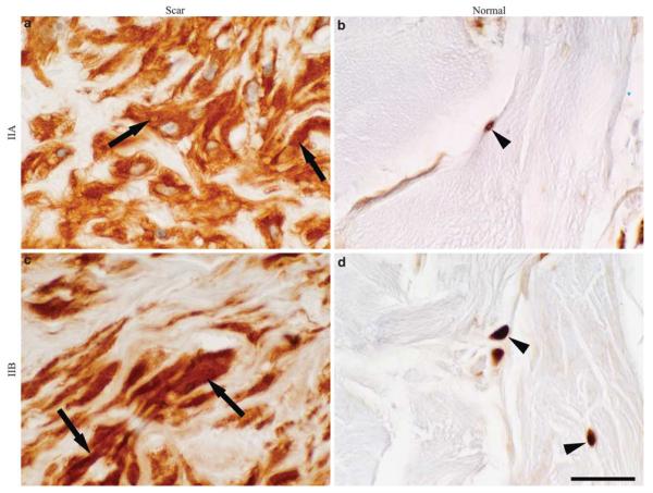
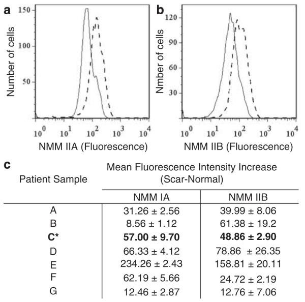
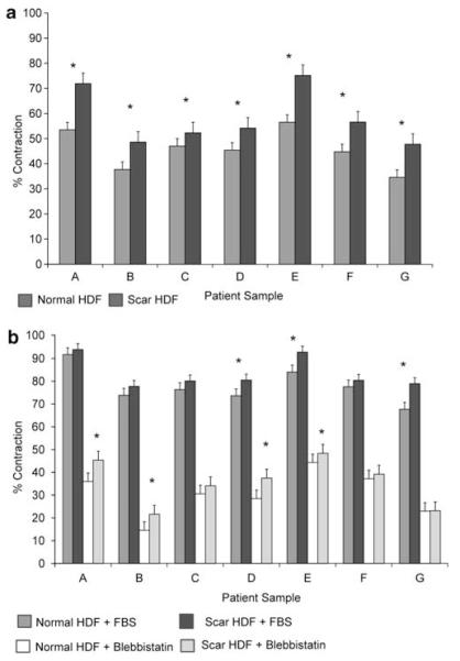
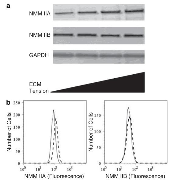
Scar contracture is believed to be caused by the cell contractility during the remodeling phase of wound healing. Cell contractility is mediated by non-muscle myosin II (NMMII) and actin, but the temporal-spatial expression profile of NMMII isoforms A and B (IIA and IIB) during the remodeling phase and the role of NMMII in scar fibroblast tissue remodeling are unknown. Human scar tissue immunostained for IIA and IIB showed that both isoforms were highly expressed in scar tissue throughout the remodeling phase of repair and expression levels returned to normal after the remodeling phase. Human scar tissue immunostained for β-, γ- and α-smooth muscle actin showed that all isoforms were consistently expressed throughout the remodeling phase of repair. The β- and γ-smooth muscle actin were widely expressed throughout the dermis, but α-smooth muscle actin was only locally expressed within the dermis. In vitro, fibroblasts explanted from scar tissue were shown to express more IIA than fibroblasts explanted from normal tissue and scar fibroblasts contracted collagen lattices to a greater extent than normal fibroblasts. Blebbistatin was used to demonstrate the function of NMMII in collagen lattice contraction. In normal tissue, fibroblasts are stress-shielded from external tensile stress by the extracellular matrix. After dermal injury and during remodeling, fibroblasts are exposed to a matrix of increased stiffness. The effect of matrix stiffness on IIA and IIB expression was examined. IIA expression was greater in fibroblasts cultured in collagen lattices with increasing stiffness, and in fibroblasts cultured on glass slides compared with polyacrylamide gels with stiffness of 1 kPa. In conclusion, NMMII and actin isoform expression changes coordinately with the remodeling phase of repair, and NMMII is increased as matrix stiffness increases. As NMMII expression increases, so does the fibroblast contractility.
Figures
Similar articles
-
The different roles of myosin IIA and myosin IIB in contraction of 3D collagen matrices by human fibroblasts.Exp Cell Res. 2014 Aug 15;326(2):295-306. doi: 10.1016/j.yexcr.2014.04.013. Epub 2014 Apr 25. Exp Cell Res. 2014. PMID: 24768700 Free PMC article.
-
Dupuytren's fibroblast contractility by sphingosine-1-phosphate is mediated through non-muscle myosin II.J Hand Surg Am. 2010 Oct;35(10):1580-8. doi: 10.1016/j.jhsa.2010.07.009. J Hand Surg Am. 2010. PMID: 20888494 Free PMC article.
-
Nonmuscle Myosin II Activation Regulates Cell Proliferation, Cell Contraction, and Myofibroblast Differentiation in Keloid-Derived Fibroblasts.Adv Wound Care (New Rochelle). 2020 Sep;9(9):491-501. doi: 10.1089/wound.2019.0944. Epub 2020 Jan 14. Adv Wound Care (New Rochelle). 2020. PMID: 32941122 Free PMC article.
-
Myosin II isoforms in smooth muscle: heterogeneity and function.Am J Physiol Cell Physiol. 2007 Aug;293(2):C493-508. doi: 10.1152/ajpcell.00131.2007. Epub 2007 May 2. Am J Physiol Cell Physiol. 2007. PMID: 17475667 Review.
-
Mammalian nonmuscle myosin II comes in three flavors.Biochem Biophys Res Commun. 2018 Nov 25;506(2):394-402. doi: 10.1016/j.bbrc.2018.03.103. Epub 2018 Mar 17. Biochem Biophys Res Commun. 2018. PMID: 29550471 Free PMC article. Review.
Cited by
-
Inhibition of Hsp90 Counteracts the Established Experimental Dermal Fibrosis Induced by Bleomycin.Biomedicines. 2021 Jun 7;9(6):650. doi: 10.3390/biomedicines9060650. Biomedicines. 2021. PMID: 34200311 Free PMC article.
-
Mechanotransduction in Skin Inflammation.Cells. 2022 Jun 25;11(13):2026. doi: 10.3390/cells11132026. Cells. 2022. PMID: 35805110 Free PMC article. Review.
-
Human mesenchymal stem cells express a myofibroblastic phenotype in vitro: comparison to human cardiac myofibroblasts.Mol Cell Biochem. 2014 Jul;392(1-2):187-204. doi: 10.1007/s11010-014-2030-6. Epub 2014 Apr 2. Mol Cell Biochem. 2014. PMID: 24691634
-
Ixodes scapularis Tick Saliva Proteins Sequentially Secreted Every 24 h during Blood Feeding.PLoS Negl Trop Dis. 2016 Jan 11;10(1):e0004323. doi: 10.1371/journal.pntd.0004323. eCollection 2016 Jan. PLoS Negl Trop Dis. 2016. PMID: 26751078 Free PMC article.
-
Unraveling SSc Pathophysiology; The Myofibroblast.Front Immunol. 2018 Nov 13;9:2452. doi: 10.3389/fimmu.2018.02452. eCollection 2018. Front Immunol. 2018. PMID: 30483246 Free PMC article. Review.
References
-
- Renovo. Renovo Ltd. [accessed 13th Oct 2010];Biotechanalytics. 2001 http://www.renovo.com/content.asp?c_id=16.
-
- Nedelec B, Ghahary A, Scott PG, et al. Control of wound contraction. Basic and clinical features. Hand Clin. 2000;16:289–302. - PubMed
-
- Stadelmann WK, Digenis AG, Tobin GR. Physiology and healing dynamics of chronic cutaneous wounds. Am J Surg. 1998;176(2A Suppl):26S–38S. - PubMed
-
- Clark RA. Cutaneous tissue repair: basic biologic considerations. I. J Am Acad Dermatol. 1985;13(5 Part 1):701–725. - PubMed
-
- Hanley WS, Snyder ME, Field L, et al. Dose-response studies of penicillamine and related compounds in reducing skin-tensile strength of rats. Chem Biol Interact. 1978;21:263–270. - PubMed
Publication types
MeSH terms
Substances
Grants and funding
LinkOut - more resources
Full Text Sources
Other Literature Sources
Medical
Molecular Biology Databases
Research Materials
