. 2011 Mar 31;30(13):1531-41.
doi: 10.1038/onc.2010.533. Epub 2010 Nov 22.

Ribosomal protein S6 is highly expressed in non-Hodgkin lymphoma and associates with mRNA containing a 5' terminal oligopyrimidine tract

Affiliations

Ribosomal protein S6 is highly expressed in non-Hodgkin lymphoma and associates with mRNA containing a 5' terminal oligopyrimidine tract

P R Hagner et al. Oncogene. .

Abstract

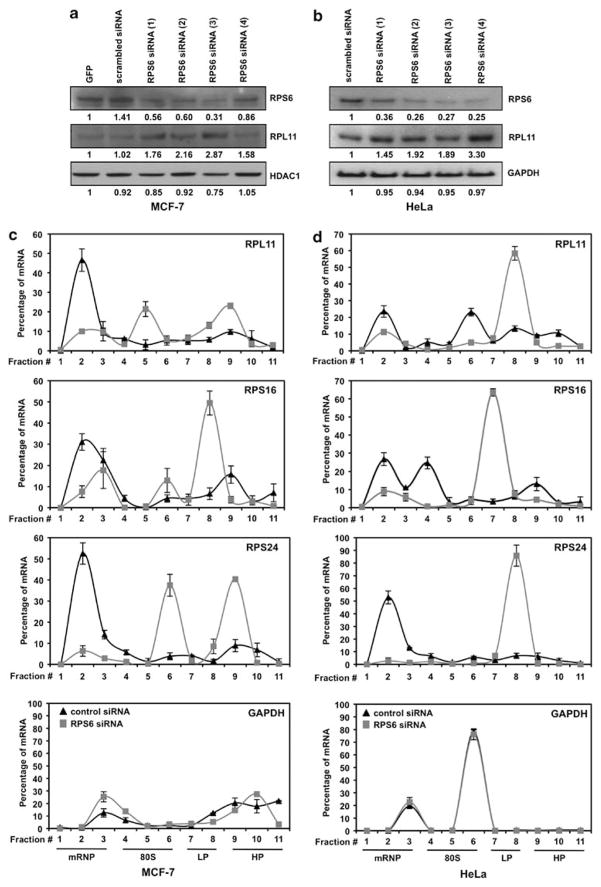
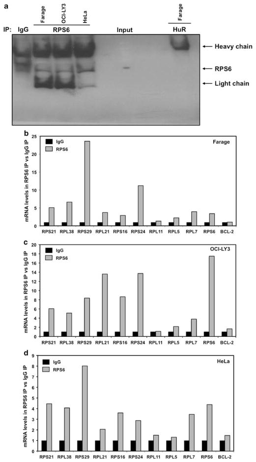
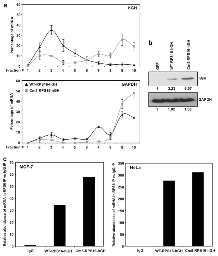
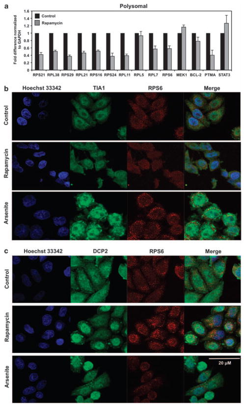
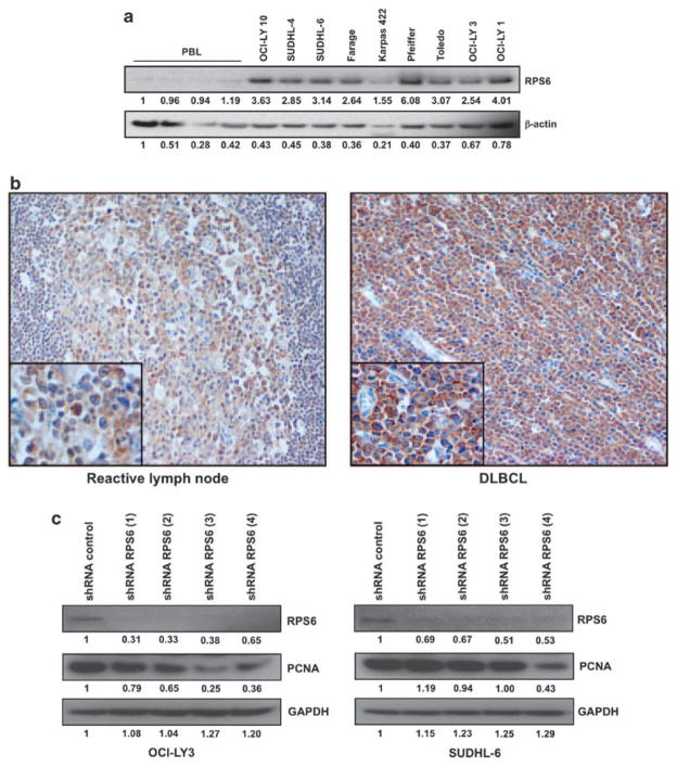
The molecular mechanism(s) linking tumorigenesis and morphological alterations in the nucleolus are presently coming into focus. The nucleolus is the cellular organelle in which the formation of ribosomal subunits occurs. Ribosomal biogenesis occurs through the transcription of ribosomal RNA (rRNA), rRNA processing and production of ribosomal proteins. An error in any of these processes may lead to deregulated cellular translation, evident in multiple cancers and 'ribosomopathies'. Deregulated protein synthesis may be achieved through the overexpression of ribosomal proteins as seen in primary leukemic blasts with elevated levels of ribosomal proteins S11 and S14. In this study, we demonstrate that ribosomal protein S6 (RPS6) is highly expressed in primary diffuse large B-cell lymphoma (DLBCL) samples. Genetic modulation of RPS6 protein levels with specifically targeted short hairpin RNA (shRNA) lentiviruses led to a decrease in the actively proliferating population of cells compared with control shRNA. Low-dose rapamycin treatments have been shown to affect the translation of 5' terminal oligopyrimidine (5' TOP) tract mRNA, which encodes the translational machinery, implicating RPS6 in 5' TOP translation. Recently, it was shown that disruption of 40S ribosomal biogenesis through specific small inhibitory RNA knockdown of RPS6 defined RPS6 as a critical regulator of 5' TOP translation. For the first time, we show that RPS6 associates with multiple mRNAs containing a 5' TOP tract. These findings expand our understanding of the mechanism(s) involved in ribosomal biogenesis and deregulated protein synthesis in DLBCL.


Conflict of interest statement

Conflict of interest

The authors declare no conflict of interest.

Figures

Figure 1
Figure 1
Specific siRNA knockdown of RPS6 leads to an increased translation of mRNA containing a 5′ TOP motif. (a, b) Representative western blot analysis of MCF-7 or HeLa cells 48 h after transfection with control and RPS6 siRNA. A volume of 30 μg of total protein lysates was loaded and the abundance of RPS6, RPL11, HDAC1 and GAPDH was assessed. (c, d) Cytoplasmic lysates from either scrambled siRNA or RPS6 siRNA cells were fractionated through sucrose gradient centrifugation. Quantitative PCR was performed on the RNA isolated from each fraction using specific primers for RPL11, RPS16, RPS24 and GAPDH. Fold changes were calculated for target mRNA in each fraction and summed together to define a total mRNA population. Signal from each fraction was divided by the total population to calculate the percentage of mRNA present per fraction from either scrambled siRNA or RPS6 siRNA cells. Graphs represent the mean and standard deviation from three independent assays. Cytoplasmic fractions may be grouped together as free messenger ribonucleoprotein (mRNP), 80S ribosomes, and light (LP) or heavy (HP) polysomes. GAPDH, glyceraldehyde 3-phosphate dehydrogenase; GFP, green florescent protein.
Figure 2
Figure 2
RPS6 associates with multiple 5′ TOP messages. (a) Representative immunoprecipitation assays performed as described (Materials and methods) but followed by western blot analysis to assess the abundance of RPS6 protein in the IP material, isotype IgG and non-RPS6-binding protein HuR were included as controls. (b–d) Cytoplasmic lysates from Farage, OCI-LY3 and HeLa cell lines were used for immunoprecipitation assays, using anti-RPS6 antibody. RNA was isolated and reverse transcription followed by qPCR was performed to measure abundance of RNA. Graph represents the mean from three independent assays.
Figure 3
Figure 3
RPS6 associates with both wild-type and mutant 5′ TOP messages. (a) Quantitative PCR analysis of the distribution on polysomes of hGH reporter mRNA in HeLa cells transfected with either WT-RPS16-hGH or Cm5-RPS16-hGH plasmids (b) Representative western blot analysis of HeLa cells transfected with either control green florescent protein (GFP), WT-RPS16-hGH or Cm5-RPS16-hGH. A volume of 10 μg of total protein lysates was loaded and the abundance of hGH and glyceraldehyde 3-phosphate dehydrogenase (GAPDH) was assessed. (c) Cytoplasmic lysates from MCF-7 and HeLa cell lines were used for immunoprecipitation assays, using anti-RPS6 antibody. RNA was isolated and reverse transcription followed by qPCR was performed to measure abundance of RNA. Graph represents the mean from three independent assays.
Figure 4
Figure 4
RPS6 does not associate with either stress granules or processing bodies after rapamycin treatment. (a) Relative association of mRNA with actively translating polysomes was tested by preparing cytoplasmic lysates from HeLa cells treated with either dimethyl sulfoxide or 10 nM rapamycin for 8 h, fractionating them through sucrose gradients and collecting 11 fractions for analysis. mRNA levels from pooled fractions (8–11) comprised of translating RNA were quantified by real-time qPCR and normalized to glyceraldehyde 3-phosphate dehydrogenase (GAPDH) mRNA. At the times indicated following addition of rapamycin or after a 60-min incubation with 0.5 mM sodium arsenite, the subcellular localizations of (b) RPS6 (red) and TIA-1 (green) or (c) RPS6 (red) and DCP2 (green) were monitored by immunofluorescence. Merge: yellow indicates colocalization of RPS6 and TIA-1 or DCP2 signals.
Figure 5
Figure 5
RPS6 is overexpressed in DLBCL and shRNA knockdown of RPS6 resulted in decreased proliferation. (a) Representative western blot analysis of peripheral blood lymphocytes from multiple patients and several DLBCL cell lines. A volume of 30 μg of total protein lysates was loaded and the abundance of RPS6 and β-actin was assessed. (b) Representative immunohistochemistry staining of paraffin embedded reactive lymph node or primary DLBCL lymph node with anti-RPS6 antibody (original magnifications of × 200 and × 400). (c) Representative western blot analysis of OCI-LY3 and SUDHL-6 cells transduced with multiple shRNA sequences. A volume of 30 μg of total protein lysates was loaded and the abundance of RPS6, PCNA and GAPDH was assessed. GAPDH, glyceraldehyde 3-phosphate dehydrogenase; PBL, peripheral blood lymphocyte; PCNA, proliferating cell nuclear antigen.

References

    1. Avni D, Biberman Y, Meyuhas O. The 5′ terminal oligopyrimidine tract confers translational control on TOP mRNAs in a cell type- and sequence context-dependent manner. Nucleic Acids Res. 1997;25:995–1001. - PMC - PubMed
    1. Badis G, Saveanu C, Fromont-Racine M, Jacquier A. Targeted mRNA degradation by deadenylation-independent decapping. Mol Cell. 2004;15:5–15. - PubMed
    1. Dai B, Kim O, Xie Y, Guo Z, Xu K, Wang B, et al. Tyrosine kinase Etk/BMX is up-regulated in human prostate cancer and its overexpression induces prostate intraepithelial neoplasia in mouse. Cancer Res. 2006;66:8058–8064. - PubMed
    1. Draptchinskaia N, Gustavsson P, Andersson B, Pettersson M, Willig TN, Dianzani I, et al. The gene encoding ribosomal protein S19 is mutated in Diamond-Blackfan anaemia. Nat Genet. 1999;21:169–175. - PubMed
    1. Espinosa L, Martín M, Nicolas A, Fabre M, Navarro E. Primary sequence of the human, lysine-rich, ribosomal protein RPL38 and detection of an unusual RPL38 processed pseudogene in the promoter region of the type-1 angiotensin II receptor gene. Biochim Biophys Acta. 1997;1354:58–64. - PubMed

Publication types

MeSH terms