. 2011 Jan 5;30(1):104-14.
doi: 10.1038/emboj.2010.291. Epub 2010 Nov 23.

RsgA releases RbfA from 30S ribosome during a late stage of ribosome biosynthesis

Affiliations

RsgA releases RbfA from 30S ribosome during a late stage of ribosome biosynthesis

Simon Goto et al. EMBO J. .

Abstract

RsgA is a 30S ribosomal subunit-binding GTPase with an unknown function, shortage of which impairs maturation of the 30S subunit. We identified multiple gain-of-function mutants of Escherichia coli rbfA, the gene for a ribosome-binding factor, that suppress defects in growth and maturation of the 30S subunit of an rsgA-null strain. These mutations promote spontaneous release of RbfA from the 30S subunit, indicating that cellular disorders upon depletion of RsgA are due to prolonged retention of RbfA on the 30S subunit. We also found that RsgA enhances release of RbfA from the mature 30S subunit in a GTP-dependent manner but not from a precursor form of the 30S subunit. These findings indicate that the function of RsgA is to release RbfA from the 30S subunit during a late stage of ribosome biosynthesis. This is the first example of the action of a GTPase on the bacterial ribosome assembly described at the molecular level.


Conflict of interest statement

The authors declare that they have no conflict of interest.

Figures

Figure 1
Figure 1
Mutations in rbfA suppress disorders of rsgA cells. (AC) Mutations in rbfA restore cell growth. Panels represent growth of (A) W3110, W3110ΔrsgA and W3110ΔrsgA-derived mutant strains, (B) W3110ΔrsgA strains harbouring plasmids carrying mutant rbfA and (C) W3110ΔrbfA strains harbouring plasmids carrying mutant rbfA. The effects of only two mutations are shown, and those of the remaining mutations are shown in Supplementary Figure 2. Doubling times were 0.9 h for W3110, 1.1–1.4 h for W3110ΔrsgA-derived mutant strains and 2.0 h for W3110ΔrsgA, W3110ΔrbfA and W3110ΔrsgAΔrbfA. (D, E) Mutations in rbfA restore 70S ribosomes to an almost normal level. The lysate of 10 mg (wet weight) of cells was subjected to a sucrose density gradient centrifugation. The direction of sedimentation is right to left. Panels represent ribosome profiles of (D) W3110, W3110ΔrsgA, W3110ΔrbfA and W3110ΔrsgAΔrbfA, and (E) growth-restored mutants derived from W3110ΔrsgA. (F) Mutations in rbfA suppress accumulation of 17S RNA. Total RNA from log-phase cells of each strain was analysed by 1.5% agarose gel electrophoresis. The bands of 23S, 17S and 16S RNAs are indicated with arrows.
Figure 2
Figure 2
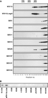
Localization of RbfA in cells of wild-type, rsgA and rsgA-suppressing mutants detected by anti-RbfA antibody. (A) Localization of RbfA in the 30S fraction and other fractions. The lysate of 100 mg (wet weight) of log-phase cells was subjected to a sucrose density gradient centrifugation and divided into 20 fractions after sedimentation. One-third of each fraction was precipitated with acetone and separated by SDS–PAGE. RbfA was immunochemically detected. The 70S, 50S and 30S fractions of sedimentations are indicated. (B) RbfA in the 30S fraction. The lysate of 10 mg (wet weight) of cells was subjected to a sucrose density gradient centrifugation. 0.15 A260 units of the 30S fraction was precipitated with acetone followed by SDS–PAGE. RbfA was detected by western immunoblotting.
Figure 3
Figure 3
RbfA promotes association of RsgA with the 30S subunit. (A) Hydrolysis of GTP (2.5 mM) by RsgA (300 nM) with or without the 30S subunits (50 nM) was monitored with increasing concentrations of RbfA. The ratio of the initial velocity (V0) to that without RbfA is indicated by the right vertical axis. The experiment was repeated three times and the average values with s.d. are presented. (B) Hydrolysis of GTP (2.5 mM) by RsgA (300 nM) with or without RbfA (six-fold concentration each of that of the 30S subunits) was monitored with increasing concentrations of the 30S subunits. The ratio of the initial velocity (V0) to that with the 30S subunits (50 nM) in the absence of RbfA is indicated by the right vertical axis. The experiment was repeated three times and the average values with s.d. are presented. P-values calculated by unpaired t-test with Welch correction between V0 with and without RbfA were 0.02, 0.006, 0.1, 0.45 and 0.86 at 50, 150, 450, 1350 and 4050 nM of the 30S subunits, respectively. (C) Complex formation between RsgA and the 30S subunit monitored by FCS. Two nM of RsgA randomly labelled with TAMRA (RsgA*) was incubated for 60 min at room temperature with 1 mM GDPNP and various concentrations of the 30S subunits in the presence or absence of RbfA (10-fold concentration each of that of the 30S subunits). Diffusion time measured using an MF20 system (Olympus) was plotted against concentration of the 30S subunits. Diffusion times of control reactions with RsgA* and various concentrations of RbfA in the absence of the 30S subunits were also measured. The measurement was repeated five times and the average value with s.d. is presented. The difference in diffusion time of RsgA in the presence of the 30S subunits with versus without RbfA was significant at 1–16 nM of the 30S subunits (P<0.004; unpaired t-test with Welch correction).
Figure 4
Figure 4
Dissociation of RbfA from the 30S subunit in vitro. The 30S subunits (50 nM) with an excess of RbfA (500 nM) were incubated for 30 min at 37°C. The mixture was subjected to centrifugal ultrafiltration using Microcon YM-100 (Millipore) to obtain the complex of RbfA and the 30S subunits. The complex of RbfA and 30S subunits was incubated under indicated conditions. After incubation, RbfA that remained on the 30S subunits was obtained by ultrafiltration using YM-100, followed by precipitation with acetone. The precipitate was separated by SDS–PAGE and RbfA was immunochemically detected. (A) RsgA dissociates RbfA from the 30S subunits. The complex of RbfA with the 30S subunits was incubated for 30 min at 37°C with or without 500 nM of wild-type or T240A mutant of RsgA in the presence or absence of GTP or ATP. (B) Enzymatic dissociation of RbfA from the 30S subunits by RsgA. The complex of the 30S subunit and RbfA was incubated with 5 nM, 50 nM or 500 nM of RsgA in the absence (−) or presence of guanine nucleotide (GDP, GTP or GDPNP) for 30 min at 37°C.
Figure 5
Figure 5
Preference for mature or immature 30S subunit by RsgA or RbfA. (A) Complex formation between RsgA and mature or immature 30S subunits monitored by FCS. Two nM of TAMRA-labelled RsgA (RsgA*) with 1 mM GDPNP incubated for 60 min at room temperature with various concentrations of the 30S subunits in the presence or absence of RbfA. Diffusion time was measured using an MF20 and is plotted against concentration of the 30S subunits. The measurement was repeated five times and the average value with s.d. is presented. (B) Complex formation between RbfA and mature or immature 30S subunit monitored by FCS. Two nM of TAMRA-labelled RbfA (RbfA*) was incubated for 60 min at room temperature with various concentrations of the 30S subunits. Diffusion time was measured using an MF20 and is plotted against concentration of the 30S subunits. The measurement was repeated five times and the average value with standard deviation is presented. (C) Stability of the complex of RbfA and mature or immature 30S subunit. A measure of 50 nM of mature or immature 30S subunits was incubated with RbfA (500 nM) for 37°C. Washed complex of RbfA and mature or immature 30S subunit retained by ultrafiltration using YM-100 was diluted to 50 nM and incubated for 30, 60 or 120 min in the presence or absence of RsgA (50 nM) with GTP (1 mM) at 37°C. After incubation, ribosome-bound RbfA retained by ultrafiltration using YM-100 was separated by SDS–PAGE and immunochemically detected.
Figure 6
Figure 6
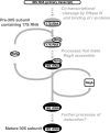
Schematic diagram of the involvement of RbfA and RsgA in the course of ribosome biosynthesis. Arrows with solid lines represent the processes established by this study. Arrows with dashed lines represent presumable processes.

Similar articles

Cited by

References

    1. Absalon C, Hamze K, Blanot D, Frehel C, Carballido-Lopez R, Holland BI, Heijenoort Jv, Séror SJ (2008) The GTPase CpgA is implicated in the deposition of the peptidoglycan sacculus in Bacillus subtilis. J Bacteriol 190: 3786–3790 - PMC - PubMed
    1. Arigoni F, Talabot F, Peitsch M, Edgerton MD, Meldrum E, Allet E, Fish R, Jamotte T, Curchod ML, Loferer H (1998) A genome-based approach for the identification of essential bacterial genes. Nat Biotechnol 16: 851–856 - PubMed
    1. Britton RA (2009) Role of GTPases in bacterial ribosome assembly. Annu Rev Microbiol 63: 155–176 - PubMed
    1. Bylund GO, Lövgren JM, Wikström PM (2001) Characterization of mutations in the metY-nusA-infB operon that suppress the slow growth of a ΔrimM mutant. J Bacteriol 183: 6095–6106 - PMC - PubMed
    1. Bylund GO, Wipemo LC, Lundberg LA, Wikström PM (1998) RimM and RbfA are essential for efficient processing of 16S rRNA in Escherichia coli. J Bacteriol 180: 73–82 - PMC - PubMed

Publication types

MeSH terms

LinkOut - more resources