Review
. 2010 Dec 15;29(24):4070-82.
doi: 10.1038/emboj.2010.294. Epub 2010 Nov 23.

Kinetochore-microtubule interactions: steps towards bi-orientation

Affiliations
Review

Kinetochore-microtubule interactions: steps towards bi-orientation

Tomoyuki U Tanaka. EMBO J. .

Abstract

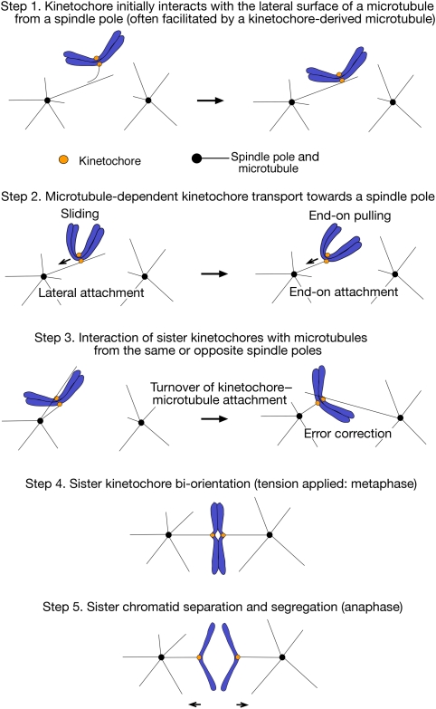
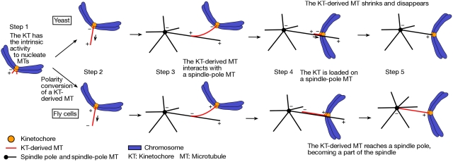
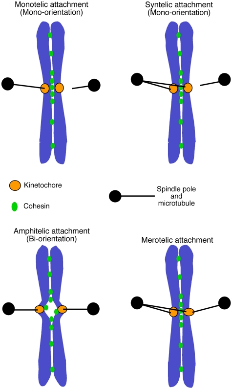
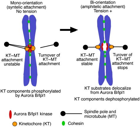
Eukaryotic cells segregate their chromosomes accurately to opposite poles during mitosis, which is necessary for maintenance of their genetic integrity. This process mainly relies on the forces generated by kinetochore-microtubule (KT-MT) attachment. During prometaphase, the KT initially interacts with a single MT extending from a spindle pole and then moves towards a spindle pole. Subsequently, MTs from the other spindle pole also interact with the KT. Eventually, one sister KT becomes attached to MTs from one pole while the other sister to those from the other pole (sister KT bi-orientation). If sister KTs interact with MTs with aberrant orientation, this must be corrected to attain proper bi-orientation (error correction) before the anaphase is initiated. Here, I discuss how KTs initially interact with MTs and how this interaction develops into bi-orientation; both processes are fundamentally crucial for proper chromosome segregation in the subsequent anaphase.


Conflict of interest statement

The author declares that he has no conflict of interest.

Figures

Figure 1
Figure 1
Step-wise development of kinetochore–microtubule interactions in eukaryotic cells. The figure shows kinetochore–microtubule (KT–MT) interactions during prometaphase (steps 1–3), metaphase (step 4) and anaphase (step 5). Prometaphase in budding yeast is as defined in Kitamura et al (2007). Step 1: The KT initially interacts with the lateral surface of a single MT (lateral attachment), which extends from one spindle pole (spindle-pole MT) (right) (Hayden et al, 1990; Rieder and Alexander, 1990; Tanaka et al, 2005a). This process is often facilitated by the interaction between a KT-derived MT and a spindle-pole MT (left) (see Figure 2) (Khodjakov et al, 2003; Maiato et al, 2004; Kitamura et al, 2010). It is not yet known whether both or only one of the sister KTs attaches to an MT during lateral attachment. Step 2: Once loaded on a spindle-pole MT, the KT is transported along the MT lateral surface towards a spindle pole (sliding; left) (Hayden et al, 1990; Rieder and Alexander, 1990; Tanaka et al, 2005a). Subsequently, at least in yeast, the KT is tethered at the end of a single spindle-pole MT (end-on attachment) and transported further as the MT shrinks (end-on pulling; right) (Kitamura et al, 2007; Tanaka et al, 2007). Step 3: Following the KT transport towards a spindle pole, both sister KTs could attach to MTs. If both sister KTs attach to MTs from the same spindle pole (syntelic attachment; see Figure 4), the KT–MT attachment must be turned over until proper bi-orientation is established (error correction; see Figure 5) (Nicklas, 1997; Tanaka et al, 2002). Step 4: The turnover of the KT–MT attachment stops once tension is generated across sister KTs upon the establishment of bi-orientation (see Figure 5) (Nicklas, 1997; Tanaka et al, 2002; Dewar et al, 2004). The number of MTs whose plus ends attach to a single KT increases as more tension is applied on metazoan KTs (King and Nicklas, 2000), while only a single MT attaches to each sister KT in budding yeast (Winey et al, 1995). Step 5: Once all sister KTs bi-orient on the spindle, cohesion between sister chromatids is removed, causing sister chromatid segregation to opposite spindle poles during anaphase (Nasmyth, 2002). Sister chromatid separation proceeds along a chromosome arm from the centromere to the telomere in budding yeast (Renshaw et al, 2010). The end-on KT–MT attachment is maintained as the MTs depolymerize and sister KTs are pulled towards the opposite poles during anaphase A, which is followed by the pole-to-pole distance being enlarged during anaphase B.
Figure 2
Figure 2
Kinetochore-derived microtubules in yeast and fly cells. The figure shows generation of MTs at KTs and the roles of KT-derived MTs during early mitosis in yeast (budding yeast) and fly (Drosophila) cells. Step 1: In both organisms, the KT can nucleate MTs and generate short MTs with distal plus ends (see discussion in Maiato et al, 2004; Kitamura et al, 2010); ‘+' and ‘−' designate the polarity of relevant MTs. Step 2: In yeast (top), a KT-derived MT extends by tubulin polymerization at the MT plus end, which remains distal to the KT (Kitamura et al, 2010). On the other hand, in fly cells (bottom), the plus end of a KT-derived MT is caught on the KT (polarity conversion) and this MT extends by tubulin polymerization at the KT, with the minus end distal to the KT (Khodjakov et al, 2003; Maiato et al, 2004). Step 3: In both organisms, the KT-derived MT interacts with a spindle-pole MT along their length, which facilitates KT loading onto the lateral surface of the spindle-pole MT (Khodjakov et al, 2003; Kitamura et al, 2010). Steps 4 and 5: Once the KT is loaded onto the spindle-pole MTs in yeast, already-existing KT-derived MTs shrink and disappear, and the KT cannot generate new MTs (Kitamura et al, 2010). On the other hand, in fly cells, KT-derived MTs can extend further and often reach a spindle pole, becoming a part of the spindle (Khodjakov et al, 2003; Maiato et al, 2004).
Figure 3
Figure 3
The Ndc80 and Dam1 complexes make interface for kinetochore–microtubule attachment. (A) Structure of the Ndc80 complex, which consists of Ndc80, Nuf2, Spc24 and Spc25 proteins (Wei et al, 2007; Ciferri et al, 2008). The globular domains of Ndc80 and Nuf2 are folded into calponin-homology (CH) domains. (B) The role of the Ndc80 and Dam1 complexes in the conversion of lateral to end-on KT–MT attachment (see Figure 1, step 2). During the lateral attachment, the Ndc80 complex (shown in red) binds an MT at its CH domains and unstructured N-terminus (Cheeseman et al, 2006; Wei et al, 2007; Ciferri et al, 2008). To convert lateral to end-on attachment, it is probably crucial that the Ndc80 complex becomes associated with the Dam1 complex (shown in green) (Lampert et al, 2010; Tien et al, 2010; our unpublished results), which localizes at the MT plus end and forms an oligomer and/or a ring encircling the MT (reviewed in Westermann et al, 2007; Nogales and Ramey, 2009). The Ndc80 N-terminus is suggested to strengthen the MT association (Guimaraes et al, 2008; Miller et al, 2008). This region may be also important for inter-complex association (Ciferri et al, 2008; Alushin et al, 2010). It is not yet clear whether the Ndc80 complexes go through the inside or outside of a Dam1-complex ring, if the ring is formed around an MT.
Figure 4
Figure 4
Modes of kinetochore–microtubule interactions. Monotelic attachment: one of the sister KTs attaches to MTs, whereas the other does not attach to any MTs. Syntelic attachment: both sister KTs attach to MTs extending from one spindle pole. As a result of monotelic or syntelic attachment, sister KTs ‘mono-orient'; that is, they are connected to only one spindle pole directly or indirectly. Amphitelic attachment: one sister kinetochore becomes attached to microtubules from one pole while the other sister to those from the other pole. As a result of amphitelic attachment, sister KTs ‘bi-orient'; that is they are connected to the opposite spindle poles. Merotelic attachment: one sister KT simultaneously attaches to MTs extending from both spindle poles. The figure is adapted from a figure in Tanaka et al (2005b).
Figure 5
Figure 5
How Aurora B/Ipl1 kinase promotes the error correction of kinetochore–microtubule attachment. The Aurora B/Ipl1 kinase facilitates sister KT bi-orientation by promoting the turnover of KT–MT attachment in a tension-dependent manner; this process is thought to take place as follows (Tanaka et al, 2002; Dewar et al, 2004; Liu et al, 2009). While syntelic attachment does not generate tension across sister KTs (left), the Aurora B/Ipl1 kinase phosphorylates KT components such as Ndc80 and Dam1 (see references in text), which promotes turnover of the KT–MT attachment. When bi-orientation is established as a result of this turnover (right), tension is applied across sister KTs, causing delocalization of the KT substrates from Aurora B/Ipl1, which leads to their de-phosphorylation. This in turn makes the KT–MT attachment more stable and stops the turnover of this attachment. Thus, sister KT bi-orientation is stably maintained and the error correction is completed for the KT–MT attachment.

Similar articles

Cited by

References

    1. Akiyoshi B, Nelson CR, Ranish JA, Biggins S (2009a) Analysis of Ipl1-mediated phosphorylation of the Ndc80 kinetochore protein in Saccharomyces cerevisiae. Genetics 183: 1591–1595 - PMC - PubMed
    1. Akiyoshi B, Nelson CR, Ranish JA, Biggins S (2009b) Quantitative proteomic analysis of purified yeast kinetochores identifies a PP1 regulatory subunit. Genes Dev 23: 2887–2899 - PMC - PubMed
    1. Alushin GM, Ramey VH, Pasqualato S, Ball DA, Grigorieff N, Musacchio A, Nogales E (2010) The Ndc80 kinetochore complex forms oligomeric arrays along microtubules. Nature 467: 805–810 - PMC - PubMed
    1. Anderson M, Haase J, Yeh E, Bloom K (2009) Function and assembly of DNA looping, clustering, and microtubule attachment complexes within a eukaryotic kinetochore. Mol Biol Cell 20: 4131–4139 - PMC - PubMed
    1. Andrews PD, Ovechkina Y, Morrice N, Wagenbach M, Duncan K, Wordeman L, Swedlow JR (2004) Aurora B regulates MCAK at the mitotic centromere. Dev Cell 6: 253–268 - PubMed

Publication types