

Efficient generation of hepatoblasts from human ES cells and iPS cells by transient overexpression of homeobox gene HEX
- PMID: 21102561
- PMCID: PMC3034848
- DOI: 10.1038/mt.2010.241
Efficient generation of hepatoblasts from human ES cells and iPS cells by transient overexpression of homeobox gene HEX
Abstract
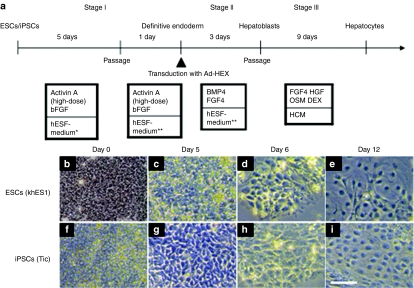
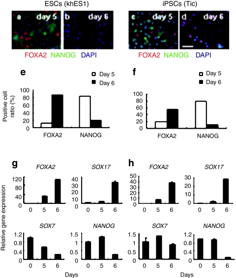
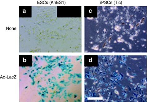
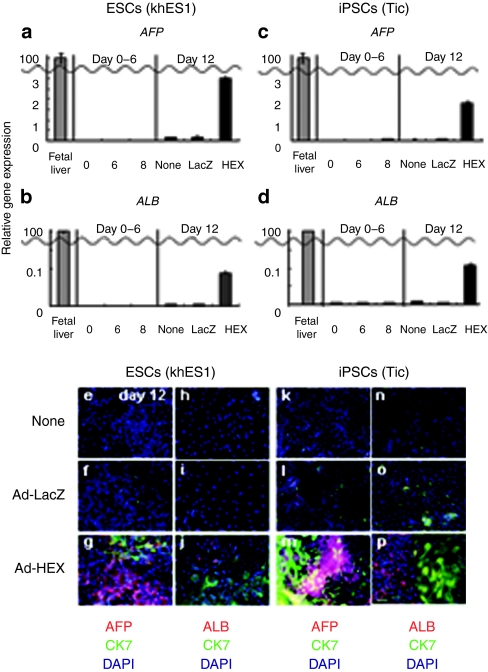
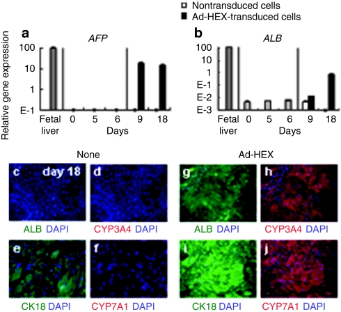
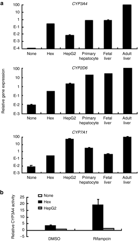
Human embryonic stem cells (ESCs) and induced pluripotent stem cells (iPSCs) have the potential to differentiate into all cell lineages, including hepatocytes, in vitro. Induced hepatocytes have a wide range of potential application in biomedical research, drug discovery, and the treatment of liver disease. However, the existing protocols for hepatic differentiation of PSCs are not very efficient. In this study, we developed an efficient method to induce hepatoblasts, which are progenitors of hepatocytes, from human ESCs and iPSCs by overexpression of the HEX gene, which is a homeotic gene and also essential for hepatic differentiation, using a HEX-expressing adenovirus (Ad) vector under serum/feeder cell-free chemically defined conditions. Ad-HEX-transduced cells expressed α-fetoprotein (AFP) at day 9 and then expressed albumin (ALB) at day 12. Furthermore, the Ad-HEX-transduced cells derived from human iPSCs also produced several cytochrome P450 (CYP) isozymes, and these P450 isozymes were capable of converting the substrates to metabolites and responding to the chemical stimulation. Our differentiation protocol using Ad vector-mediated transient HEX transduction under chemically defined conditions efficiently generates hepatoblasts from human ESCs and iPSCs. Thus, our methods would be useful for not only drug screening but also therapeutic applications.
Figures
Similar articles
-
Efficient generation of functional hepatocytes from human embryonic stem cells and induced pluripotent stem cells by HNF4α transduction.Mol Ther. 2012 Jan;20(1):127-37. doi: 10.1038/mt.2011.234. Epub 2011 Nov 8. Mol Ther. 2012. PMID: 22068426 Free PMC article.
-
Efficient generation of hepatocyte-like cells from human induced pluripotent stem cells.Cell Res. 2009 Nov;19(11):1233-42. doi: 10.1038/cr.2009.107. Epub 2009 Sep 8. Cell Res. 2009. PMID: 19736565
-
The homeobox gene Hex regulates hepatocyte differentiation from embryonic stem cell-derived endoderm.Hepatology. 2010 Feb;51(2):633-41. doi: 10.1002/hep.23293. Hepatology. 2010. PMID: 20063280
-
[Optimization of adenovirus vectors for transduction in embryonic stem cells and induced pluripotent stem cells].Yakugaku Zasshi. 2011;131(9):1333-8. doi: 10.1248/yakushi.131.1333. Yakugaku Zasshi. 2011. PMID: 21881308 Review. Japanese.
-
iPS cells: a source of cardiac regeneration.J Mol Cell Cardiol. 2011 Feb;50(2):327-32. doi: 10.1016/j.yjmcc.2010.10.026. Epub 2010 Oct 30. J Mol Cell Cardiol. 2011. PMID: 21040726 Review.
Cited by
-
Generation of functional hepatic cells from pluripotent stem cells.J Stem Cell Res Ther. 2012 Aug 15;Suppl 10(8):1-7. doi: 10.4172/2157-7633.S10-008. J Stem Cell Res Ther. 2012. PMID: 25364624 Free PMC article.
-
Induced pluripotent stem cells as a source of hepatocytes.Curr Pathobiol Rep. 2014 Mar;2(1):11-20. doi: 10.1007/s40139-013-0039-2. Curr Pathobiol Rep. 2014. PMID: 25650171 Free PMC article.
-
Efficient and directive generation of two distinct endoderm lineages from human ESCs and iPSCs by differentiation stage-specific SOX17 transduction.PLoS One. 2011;6(7):e21780. doi: 10.1371/journal.pone.0021780. Epub 2011 Jul 7. PLoS One. 2011. PMID: 21760905 Free PMC article.
-
Fine Tuning of Hepatocyte Differentiation from Human Embryonic Stem Cells: Growth Factor vs. Small Molecule-Based Approaches.Stem Cells Int. 2019 Jan 22;2019:5968236. doi: 10.1155/2019/5968236. eCollection 2019. Stem Cells Int. 2019. PMID: 30805010 Free PMC article.
-
Improved Survival and Initiation of Differentiation of Human Induced Pluripotent Stem Cells to Hepatocyte-Like Cells upon Culture in William's E Medium followed by Hepatocyte Differentiation Inducer Treatment.PLoS One. 2016 Apr 13;11(4):e0153435. doi: 10.1371/journal.pone.0153435. eCollection 2016. PLoS One. 2016. PMID: 27073925 Free PMC article.
References
-
- Thomson JA, Itskovitz-Eldor J, Shapiro SS, Waknitz MA, Swiergiel JJ, Marshall VS, et al. Embryonic stem cell lines derived from human blastocysts. Science. 1998;282:1145–1147. - PubMed
-
- Takahashi K, Tanabe K, Ohnuki M, Narita M, Ichisaka T, Tomoda K, et al. Induction of pluripotent stem cells from adult human fibroblasts by defined factors. Cell. 2007;131:861–872. - PubMed
-
- Makino H, Toyoda M, Matsumoto K, Saito H, Nishino K, Fukawatase Y, et al. Mesenchymal to embryonic incomplete transition of human cells by chimeric OCT4/3 (POU5F1) with physiological co-activator EWS. Exp Cell Res. 2009;315:2727–2740. - PubMed
-
- Nagata, TM, Yamaguchi, S, Hirano, K, Makino, H, Nishino, K, Miyagawa, Y, et al. Efficient reprogramming of human and mouse primary extra-embryonic cells to pluripotent stem cells. Genes Cells. 2009;14:1395–1404. - PubMed
-
- Lavon N., and, Benvenisty N. Study of hepatocyte differentiation using embryonic stem cells. J Cell Biochem. 2005;96:1193–1202. - PubMed
Publication types
MeSH terms
Substances
LinkOut - more resources
Full Text Sources
Other Literature Sources
Research Materials
Miscellaneous
