

Angiotensin-2 receptors (AT1-R and AT2-R), new prognostic factors for renal clear-cell carcinoma?
- PMID: 21102591
- PMCID: PMC2994218
- DOI: 10.1038/sj.bjc.6605866
Angiotensin-2 receptors (AT1-R and AT2-R), new prognostic factors for renal clear-cell carcinoma?
Abstract
Background: The growth factor Angiotensin-2 signals through Angiotensin receptor type 1 (AT1-R) in a broad range of cell types and tumours and through the type-2 receptor (AT2-R) in a more restricted group of cell types. Although numerous forms of cancer have been shown to overexpress AT1-R, expression of AT1-R and AT2-R by human renal clear-cell carcinoma (RCCC) is not well understood. In this study, the expression of both angiotensin receptors was quantified in a retrospective series of RCCC and correlated with prognostic factors.
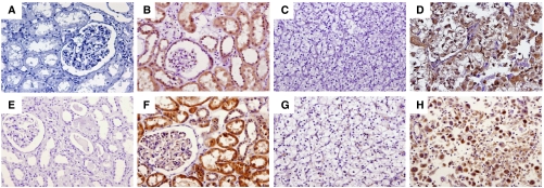
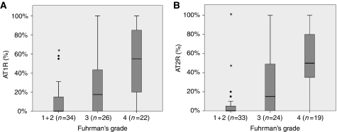
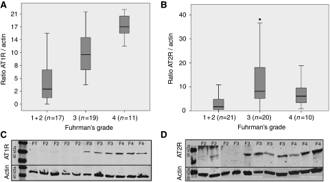
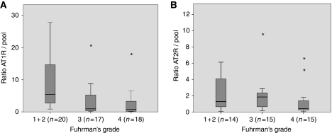
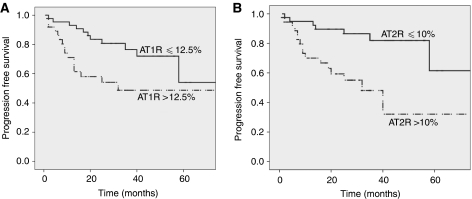
Methods: Angiotensin receptor type 1 and AT2-R expressions were quantified on tumour tissues by immunohistochemistry (IHC), western blot and quantitative reverse transcriptase PCR (qRT-PCR). IHC results were correlated to Fuhrman's grade and patient progression-free survival (PFS).
Results: A total of 84 RCCC were analysed. By IHC, AT1-R and AT2-R were expressed to a greater level in high-grade tumours (AT1-R: P<0.001, AT2-R: P<0.001). Univariate analysis showed a correlation between PFS and AT1-R or AT2-R expression (P=0.001). By multivariate analysis, only AT2-R expression correlated with PFS (HR 1.021, P=0.006) and cancer stage (P<0.001). By western blot, AT1-R and AT1-R were also found to be overexpressed in higher Fuhrman's grade (P<0.01 and P=0.001 respectively). By qRT-PCR, AT1-R but not AT2-R mRNA were downregulated (P=0.001 and P=0.118, respectively).
Conclusion: Our results show that AT1-R and AT2-R proteins are overexpressed in the most aggressive forms of RCCC and that AT2-R expression correlates with PFS. AT1-R or AT2-R blockage could, therefore, offer novel directions for anti-RCCC therapy.
Figures
Similar articles
-
Differential regulation of angiotensin II receptors during renal injury and compensatory hypertrophy in the rat.Clin Exp Pharmacol Physiol. 2005 Apr;32(4):241-8. doi: 10.1111/j.1440-1681.2005.04181.x. Clin Exp Pharmacol Physiol. 2005. PMID: 15810986
-
Expression of AT1 and AT2 angiotensin receptors in astrocytomas is associated with poor prognosis.Br J Cancer. 2008 Jul 8;99(1):160-6. doi: 10.1038/sj.bjc.6604431. Br J Cancer. 2008. PMID: 18594540 Free PMC article.
-
Renal expression of angiotensin receptors in long-term diabetes and the effects of angiotensin type 1 receptor blockade.J Hypertens. 2002 Aug;20(8):1615-24. doi: 10.1097/00004872-200208000-00025. J Hypertens. 2002. PMID: 12172324
-
[Pathophysiological function of angiotensin II AT1 and AT2 receptors and clinical application of AT1 antagonists].Nihon Rinsho. 1998 Jul;56(7):1912-8. Nihon Rinsho. 1998. PMID: 9702075 Review. Japanese.
-
AT1 and AT2 receptor in the kidney: role in health and disease.Semin Nephrol. 2004 Mar;24(2):93-100. doi: 10.1016/j.semnephrol.2003.11.009. Semin Nephrol. 2004. PMID: 15017521 Review.
Cited by
-
Expression of Components of the Renin-Angiotensin System by Cancer Stem Cells in Renal Clear Cell Carcinoma.Biomolecules. 2021 Apr 7;11(4):537. doi: 10.3390/biom11040537. Biomolecules. 2021. PMID: 33916968 Free PMC article.
-
[Advance in Research of Angiotensin II and Its Receptor and Malignant Tumor].Zhongguo Fei Ai Za Zhi. 2016 Sep 20;19(9):615-9. doi: 10.3779/j.issn.1009-3419.2016.09.10. Zhongguo Fei Ai Za Zhi. 2016. PMID: 27666553 Free PMC article. Review. Chinese.
-
GPCRs and cancer.Acta Pharmacol Sin. 2012 Mar;33(3):351-62. doi: 10.1038/aps.2011.183. Epub 2012 Jan 23. Acta Pharmacol Sin. 2012. PMID: 22266725 Free PMC article. Review.
-
Sunitinib combined with angiotensin-2 type-1 receptor antagonists induces more necrosis: a murine xenograft model of renal cell carcinoma.Biomed Res Int. 2014;2014:901371. doi: 10.1155/2014/901371. Epub 2014 May 22. Biomed Res Int. 2014. PMID: 24967411 Free PMC article.
-
Impact of Angiotensin Receptor Blocker Use on Overall Survival Among Patients Undergoing Resection for Pancreatic Cancer.World J Surg. 2017 Sep;41(9):2361-2370. doi: 10.1007/s00268-017-4021-8. World J Surg. 2017. PMID: 28429090
References
-
- Arafat HA, Gong Q, Chipitsyna G, Rizvi A, Saa CT, Yeo CJ (2007) Antihypertensives as novel antineoplastics: angiotensin-I-converting enzyme inhibitors and angiotensin II type 1 receptor blockers in pancreatic ductal adenocarcinoma. J Am Coll Surg 204: 996–1005; discussion 1005–1006 - PubMed
-
- Arendshorst WJ, Brannstrom K, Ruan X (1999) Actions of angiotensin II on the renal microvasculature. J Am Soc Nephrol 10(Suppl 11): S149–S161 - PubMed
-
- Arrieta O, Pineda-Olvera B, Guevara-Salazar P, Hernandez-Pedro N, Morales-Espinosa D, Ceron-Lizarraga TL, Gonzalez-De la Rosa CH, Rembao D, Segura-Pacheco B, Sotelo J (2008) Expression of AT1 and AT2 angiotensin receptors in astrocytomas is associated with poor prognosis. Br J Cancer 99: 160–166 - PMC - PubMed
-
- Bose SK, Gibson W, Giri S, Nath N, Donald CD (2009) Angiotensin II up-regulates PAX2 oncogene expression and activity in prostate cancer via the angiotensin II type I receptor. Prostate 69: 1334–1342 - PubMed
-
- Cao Z, Kelly DJ, Cox A, Casley D, Forbes JM, Martinello P, Dean R, Gilbert RE, Cooper ME (2000) Angiotensin type 2 receptor is expressed in the adult rat kidney and promotes cellular proliferation and apoptosis. Kidney Int 58: 2437–2451 - PubMed
Publication types
MeSH terms
Substances
LinkOut - more resources
Full Text Sources
Medical
Research Materials
