

doi: 10.1038/embor.2010.156.
Epub 2010 Nov 12.
The Ste20 kinase misshapen is essential for the invasive behaviour of ovarian epithelial cells in Drosophila
Affiliations
- PMID: 21102643
- PMCID: PMC2999858
- DOI: 10.1038/embor.2010.156
Item in Clipboard
The Ste20 kinase misshapen is essential for the invasive behaviour of ovarian epithelial cells in Drosophila
EMBO Rep.
2010 Dec.
Abstract
Stationary-to-migratory transitions of epithelial cells have a key role in development and tumour progression. Border cell migration is a powerful system in which to investigate this transition in living organisms. Here, we identify the Ste20-like kinase misshapen (msn) as a novel regulator of border-cell migration in Drosophila. Expression of msn in border cells is independent of the transcription factor slow border cells and of inputs from all pathways that are known to control border-cell migration. The msn gene functions to modulate the levels and/or distribution of Drosophila E-cadherin to promote the invasive migratory behaviour of border cells.
Conflict of interest statement
The authors declare that they have no conflict of interest.
Figures

Ovarian expression of misshapen. (A–C) Illustration of stage 8 (S8), S9 and S10 egg chambers. NCs, Oo, FCs, PCs (red) and BCs (green) are indicated. (D–F) Egg chambers from the msn06286 line stained for β-gal activity (green), the PC marker Fasciclin-III (FasIII; red) and the nuclear marker TO-PRO-3 (blue). Note that the border-cell cluster moves evenly with the main body follicle cells (empty arrowheads in E). BC, border cell; FC, follicle cell; β-gal, β-galactosidase; Fas III, Fasciclin-III; NC, nurse cell; Oo, oocyte; PC, polar cell.
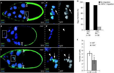
msn is required for border-cell migration. (A) WT and (B,C) msn mosaic stage 10 (S10) egg chambers. In all panels, homozygous mutant cells are labelled by the absence of GFP (green); primed letters (′) are magnifications of the white boxes. (A) At S10, posterior migration of border cells is complete. (B) An S10 msn102-mutant border-cell cluster stuck at the anterior pole of the egg chamber. The number of border cells in this msn− cluster is reduced compared with the wild type. (C) A delayed, mixed border-cell cluster containing wild-type cells at the front and msn− cells behind. (D) Quantification of the S10 migration phenotype of mutant (BC−) and mixed (BC+/−) clusters. (E) Quantification of the recruitment phenotype. Only mutant clusters were scored. An asterisk indicates a statistically significant difference (Student's test: P<0.01). Empty arrowheads, rear limit of main body follicle cells; arrowheads, mutant cells; arrow, wild-type cells. BC, border cell; GFP, green fluorescent protein; msn, misshapen; WT, wild type.
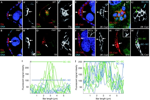
msn regulates DE-Cad levels in border cells. At stage 8 (S8), (A) WT and (B) msn− border cells localize Dlg (red) at the front, facing the germ line. (C,D) At S9, Dlg is found at high levels at PC–BC and BC–BC interfaces and low levels at the BC–NC interface in both wild-type and msn− border cells. (E,F) DE-Cad distribution in S8 egg chambers is uniform around the surface of both (E) wild-type and (F) msn− border cells. (G,H) At S10, while in wild-type S10 clusters DE-Cad is found at high levels at the PC–BC and BC–BC boundaries and at low levels at BC–NC boundaries, msn− border cells also accumulate high DE-Cad levels at BC–NC interfaces. Insets in F and H are internal controls for DE-Cad staining in follicle cells. (I,J) Histogram of relative fluorescent intensities of DE-Cad along BC–BC (green) and BC–NC (blue) boundaries, as indicated in G′ and H′. Coloured bars indicate the maximum DE-Cad fluorescence intensity found at BC–BC and BC–NC boundaries. Arrows, BC–NC contacts; arrowheads, basal side of border cells. Primed letters are single channels of corresponding merged panels. BC, border cell; DE-Cad, DE-cadherin; Dlg, Discs large; FC, follicle cell; GFP, green fluorescent protein; msn, misshapen; NC, nurse cell; PC, polar cell; WT, wild type.
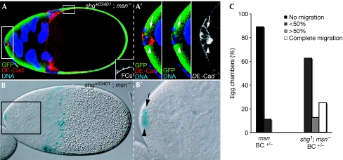
msn does not regulate DE-cadherin transcription. S11 egg chamber carrying a mixed border-cell cluster stained with anti-GFP (green) and anti-DE-Cad (red), and assayed for β-galactosidase activity. (A) DE-Cad staining in msn− cells (white arrowheads) is abnormally elevated compared with sibling wild-type controls (white arrows). The inset shows an internal control for DE-Cad staining in follicle cells. (B) msn− cells (black arrowhead) show normal β-galactosidase activity (black arrow: sibling controls). (C) Reducing DE-Cad alleviates the border-cell migration defects found in msn mixed clusters. Migration is blocked in 89% of the mixed S10 border-cell clusters and delayed in 11% (n=36). In an shg−/+ background, migration is completed in 25% of the cases and delayed in the remaining 75% (n=15). Primed letters are magnifications of the corresponding white boxes. BC, border cell; DE-Cad, Drosophila E-cadherin; FC, follicle cell; GFP, green fluorescent protein; msn, misshapen; S11, stage 11; shg, shotgun.
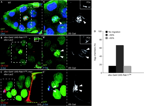
Interfering with Rab11 function affects DE-Cad levels and border-cell migration. (A) S9 controls the border-cell cluster. (B,C) S9 border cells labelled with slbo-lacZ. (B) Overexpression of UAS-Rab11DN reduces DE-Cad levels and impairs migration. (C) Overexpression of UAS-Rab11DN does not rescue the migration defects or the elevated DE-Cad levels of msn− border cells. (D) Quantification of the border-cell migration phenotype of S10 egg chambers overexpressing UAS-Rab11DN. Small insets in A and B are internal controls of DE-Cad staining. Arrowheads, BCs. Primed letters are magnifications of white boxes. BC, border cell; DE-Cad, Drosophila E-cadherin; msn, misshapen; S9, stage 9; slbo, slow border cells; WT, wild type.
References
-
- Bai J, Uehara Y, Montell DJ (2000) Regulation of invasive cell behaviours by Taiman, a Drosophila protein related to AIB1, a steroid receptor coactivator amplified in breast cancer. Cell 103: 1047–1058 - PubMed
-
- Beccari S, Teixeira L, Rorth P (2002) The JAK/STAT pathway is required for border cell migration during Drosophila oogenesis. Mech Dev 111: 115–123 - PubMed
-
- Cavalli V, Vilbois F, Corti M, Marcote MJ, Tamura K, Karin M, Arkinstall S, Gruenberg J (2001) The stress-induced MAP kinase p38 regulates endocytic trafficking via the GDI:Rab5 complex. Mol Cell 7: 421–432 - PubMed
-
- Classen AK, Anderson KI, Marois E, Eaton S (2005) Hexagonal packing of Drosophila wing epithelial cells by the planar cell polarity pathway. Dev Cell 9: 805–817 - PubMed
Publication types
MeSH terms
Substances
LinkOut - more resources
Full Text Sources
Molecular Biology Databases
Research Materials
Miscellaneous
