. 2010 Nov 12;5(11):e13952.
doi: 10.1371/journal.pone.0013952.

Sox2 is essential for formation of trophectoderm in the preimplantation embryo

Affiliations

Sox2 is essential for formation of trophectoderm in the preimplantation embryo

Maria Keramari et al. PLoS One. .

Abstract

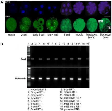
Background: In preimplantation mammalian development the transcription factor Sox2 (SRY-related HMG-box gene 2) forms a complex with Oct4 and functions in maintenance of self-renewal of the pluripotent inner cell mass (ICM). Previously it was shown that Sox2-/- embryos die soon after implantation. However, maternal Sox2 transcripts may mask an earlier phenotype. We investigated whether Sox2 is involved in controlling cell fate decisions at an earlier stage.

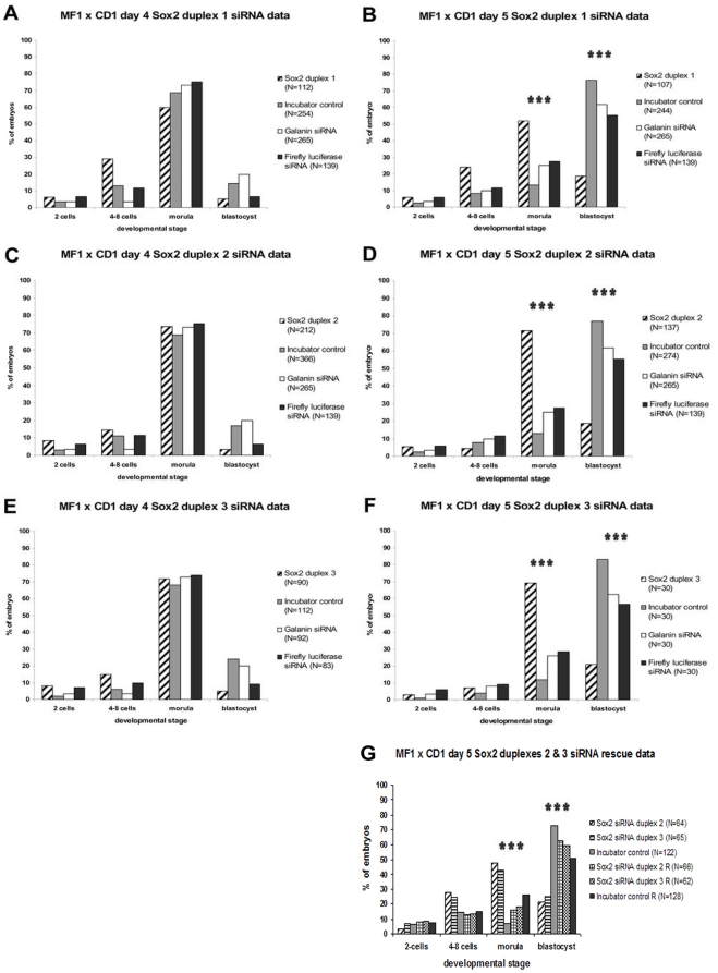
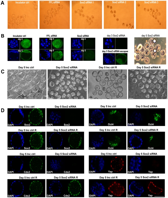
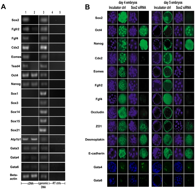
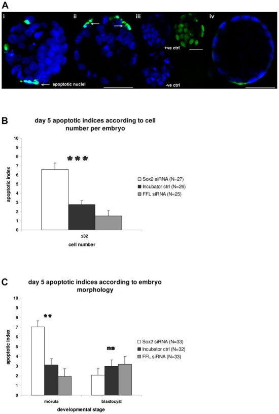
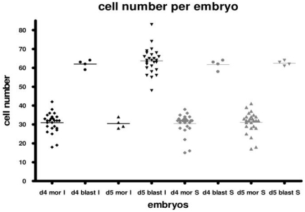
Methods and findings: We addressed the question of an earlier role for Sox2 using RNAi, which removes both maternal and embryonic Sox2 mRNA present during the preimplantation period. By depleting both maternal and embryonic Sox2 mRNA at the 2-cell stage and monitoring embryo development in vitro we show that, in the absence of Sox2, embryos arrest at the morula stage and fail to form trophectoderm (TE) or cavitate. Following knock-down of Sox2 via three different short interfering RNA (siRNA) constructs in 2-cell stage mouse embryos, we have shown that the majority of embryos (76%) arrest at the morula stage or slightly earlier and only 18.7-21% form blastocysts compared to 76.2-83% in control groups. In Sox2 siRNA-treated embryos expression of pluripotency associated markers Oct4 and Nanog remained unaffected, whereas TE associated markers Tead4, Yap, Cdx2, Eomes, Fgfr2, as well as Fgf4, were downregulated in the absence of Sox2. Apoptosis was also increased in Sox2 knock-down embryos. Rescue experiments using cell-permeant Sox2 protein resulted in increased blastocyst formation from 18.7% to 62.6% and restoration of Sox2, Oct4, Cdx2 and Yap protein levels in the rescued Sox2-siRNA blastocysts.

Conclusion and significance: We conclude that the first essential function of Sox2 in the preimplantation mouse embryo is to facilitate establishment of the trophectoderm lineage. Our findings provide a novel insight into the first differentiation event within the preimplantation embryo, namely the segregation of the ICM and TE lineages.


Conflict of interest statement

Competing Interests: The authors have declared that no competing interests exist.

Figures

Figure 1
Figure 1. Sox2 (protein and mRNA) expression in mouse preimplantation development.
Expression of Sox2 was detected at all stages (oocyte to blastocyst) of mouse preimplantation development by immunocytochemistry and RT-PCR. (A) Developmental stages of preimplantation mouse embryos immunostained for Sox2 (Abcam): single optical sections of confocal Z-series. Bar: 50 µm. (B) RT-PCR for Sox2 and β-actin in mouse preimplantation embryos, 40 cycles. Lane 1: Hyperladder IV (Bioline); 2: oocyte; 4: 2-cell; 6: 4-cell; 8: 8-cell; 10: morula; 12: blastocyst; 14: mES cells (line E14); 16: genomic DNA (Bioline). Lanes (3, 5, 7, 9, 11, 13, 15): RT –ve controls for each developmental stage.
Figure 2
Figure 2. Development of MF1xCD1 (A,B,C,D,E,F) embryos in culture after RNAi, as well as rescue phenotype of MF1XCD1 (G) embryos.
(A,B) Sox2-duplex-1-siRNA embryos (hatched, N = 112 day 4, N = 107 day 5) were compared with incubator-control (grey, N = 254 day 4, N = 244 day 5), Galanin-siRNA (white, N = 265) and FFL-siRNA (black, N = 139) embryos. Most day 4 (A) embryos formed morulae (16–32 cells), but 29% of the Sox2-duplex-1-siRNA embryos remained at the 4-8-cell stage. On day 5 (B), while 76.2% of incubator-control embryos formed blastocysts, only 18.7% of the Sox2-duplex-1-siRNA embryos reached blastocyst stage. (C,D) Sox2-duplex-2-siRNA embryos (hatched, N = 212 day 4, N = 137 day 5) were compared with incubator-control (grey, N = 366 day 4, N = 274 day 5), Galanin-siRNA (white, N = 265) and FFL-siRNA (black, N = 139) embryos. Most day 4 (C) embryos formed morulae, but 14.4% of the Sox2-duplex-2-siRNA embryos remained at the 4-8-cell stage. Only 18.7% of the day 5 (D) Sox2-duplex-2-siRNA embryos formed blastocysts. (E,F) Sox2-duplex-3-siRNA embryos (hatched, N = 90 day 4, N = 30 day 5) were compared with incubator-control (grey, N = 112 day 4, N = 30 day 5), Galanin-siRNA (white, N = 92 day 4, N = 30 day 5) and FFL-siRNA (black, N = 83 day 4, N = 30 day 5) embryos. Most day 4 (E) embryos formed morulae, while 15% of the Sox2-duplex-3-siRNA embryos remained at the 4-8-cell stage. Only 21% of the day 5 (F) Sox2-duplex-3-siRNA embryos developed to blastocysts. (G) Rescue phenotype at day 5 of development of MF1xCD1 embryos. Only 21.1% of Sox2-duplex-2-siRNA embryos (N = 64) and 25.4% of Sox2-duplex-3-siRNA embryos (N = 65) formed blastocysts. After treatment with cell-permeant Sox2 protein, 62.6% of Sox2-duplex-2-siRNA R embryos (N = 66) formed blastocysts and 59.7% of the Sox2-duplex-3-siRNA R embryos (N = 62) reached blastocyst stage. 72.75% of untreated incubator control embryos (N = 122) formed blastocysts but treatment with cell-permeant Sox2 protein (N = 128) reduced this to 50.9%. In all cases, chi-square tests revealed significant differences (p<0.0001) between the % of Sox2-siRNA, control and R morulae, as well as between the % of Sox2-siRNA, control and R blastocysts.
Figure 3
Figure 3. Sox2 expression in Sox2-siRNA and control MF1xCD1 embryos.
(A) Phase contrast images of day 5 incubator-control, FFL-siRNA, and Sox2-duplex-1, -2 and -3 siRNA embryos. While control embryos formed blastocysts, most Sox2-siRNA embryos did not. Bars: 100 µm. (B) Control and Sox2-duplex-2-siRNA embryos stained for Sox2 (Abcam) on days 4 and 5: single optical sections from confocal Z-series, DAPI-stained nuclei (blue) and Sox2 (green); bar: 50 µm. Decreased Sox2 protein expression was observed in Sox2 knock-down morulae on day 4, which persisted on day 5. Absence of Sox2 protein on day 3, as a result of the Sox2-siRNA knock-down on day 2, was also confirmed. A few blastocysts forming on day 5 within the Sox2-siRNA group (‘escapees’ from the siRNA effect) showed some Sox2-staining, although less than incubator-control blastocysts. The lethality of the Sox2-siRNA phenotype on day 5 was assessed by Trypan Blue up-take. Presence of non-viable (blue) cells was apparent in 10 out of the 12 day 5 Sox2-siRNA embryos examined. (C) Rescue phenotype in MF1xCD1 embryos: Sox2-siRNA and incubator-control embryos were cultured with and without cell-permeant Sox2 protein, and embryo development was assessed up to day 5. Phase contrast images of blastocysts from untreated day 5 incubator-control embryos (Day 5 Inc Ctrl); Lack of blastocyst formation in untreated day 5 Sox2-siRNA (duplex-2) embryos (Day 5 Sox2 siRNA); Blastocysts and some arrested embryos in day 5 control embryos treated with cell-permeant Sox2 protein (Day 5 Inc Ctrl R); Rescue phenotype on day 5 with blastocyst formation as well as some arrested embryos after treatment of Sox2-siRNA (duplex-2) embryos with cell-permeant Sox2 protein (Day 5 Sox2 siRNA R). Bars: 100 µm. (D) Comparison of D5 Sox2 siRNA embryos with untreated incubator control embryos (D5 Inc ctrl), as well as with rescued D5 Sox2 siRNA R embryos and D5 Inc ctrl R embryos for Sox2 (R&D), Oct4 (BD Biosciences), Cdx2 (Biogenex) and Yap (Cell Signaling) proteins. The images are single optical sections from confocal Z-series. Untreated D5 Inc ctrl blastocysts expressed Sox2 in ICM and TE nuclei, Oct4 in ICM nuclei, Cdx2 and Yap in TE nuclei; D5 Sox2 siRNA arrested morulae did not demonstrate Sox2, Cdx2 and Yap staining, but expressed Oct4 in inner cell nuclei; D5 Inc ctrl R blastocysts showed similar expression patterns with D5 Inc ctrl blastocysts for all 4 markers; rescued D5 Sox2 siRNA blastocysts expressed Sox2 in ICM and TE nuclei, Oct4 in ICM nuclei, Cdx2 and Yap in TE nuclei, indicating reversal of the Sox2 siRNA phenotype. At least 10 embryos were stained for each antigen and representative embryos are shown. Similar results were obtained for both Sox2 siRNA duplexes. Blue indicates DAPI stained nuclei and green/red staining for the protein of interest. In all cases immunological controls were negative (Figure S6). Bars: 100 µm.
Figure 4
Figure 4. Assessment of ICM and TE markers in Sox2-siRNA and control MF1xCD1 embryos.
(A) RT-PCR for Sox2, Fgfr2, Fgf4, Cdx2, Eomes, Tead4, Oct4, Nanog, Sox1, Sox3, Sox14, Sox15, Sox21 and beta-actin (40 cycles) on day 4: incubator-control embryos (lane 1); Sox2-duplex-2-siRNA embryos (lane 2); genomic DNA (lane 3); incubator-control morulae –RT (lane 4); Sox2-duplex-2-siRNA morulae –RT (lane 5). In the absence of Sox2 transcript after siRNA, a clear reduction of Fgfr2, Ffg4, Cdx2, Eomes and Tead4 transcripts in Sox2-siRNA embryos was observed. Oct4 and Nanog transcripts were unaffected in Sox2 knock-down morulae compared to incubator-control morulae. To ensure absence of signal did not reflect low detectable message, we performed PCR at 40 cycles. PCR for all transcripts was determined at 25, 30 and 35 cycles with results similar to those presented, but with weaker band intensity where present. Sox1, Sox3, Sox14, Sox15 and Sox21 transcripts were not detected in incubator-control morulae or Sox2 knock-down morulae. Atp1b1 (ATPase, Na+/K+ transporting, beta 1 polypeptide), Gata3, Gata4 and Gata6 transcripts were detected in comparable levels in Sox2 knock-down morulae compared to incubator-control morulae. Beta-actin transcripts were detected in both Sox2-siRNA and incubator-control embryos. This figure illustrates Sox2-duplex-2-siRNA embryo transcripts, whereas similar patterns of transcript expression for Sox2-siRNA duplexes -1 and -3 are shown in Figure S4. (B) Comparison of Sox2 knock-down embryos (as assessed by absence of fluorescence after Sox2 staining) with incubator (untreated) control embryos for ICM and TE markers. Immunofluorescence for Sox2 (Abcam), Oct4, Nanog, Cdx2 (rabbit polyclonal from Jane Collins), Eomes, Fgfr2, Fgf4, Occludin, ZO1, Desmoplakin, E-cadherin, Gata4 and Gata6 to compare protein expression between incubator control and Sox2-siRNA embryos at day 4 and 5 of preimplantation development. The images are single optical sections from confocal Z-series. Sox2-duplex-1-siRNA embryos stained for Sox2 are shown in this Figure. In all cases, after knock-down with each of the three duplexes, Sox2 siRNA embryos were negative for Sox2. For assessment of the other markers, embryos from all three knock-down construct groups were pooled. A large reduction in Sox2, Cdx2, Eomes, Fgfr2, Fgf4 and Occludin protein was observed, whereas Oct4, Nanog, ZO1, Desmoplakin, E-cadherin, Gata4 and Gata6 remained unaffected. However, Nanog staining appeared more cytoplasmic in Sox2 knock-down embryos. The selected image for the day 4 incubator control embryo stained for Cdx2, is a plane through the top of the embryo and the positively stained Cdx2 cell in the center is located in the outer layer of the embryo. The staining patterns for Sox2 and Cdx2 were confirmed with different Sox2 (Chemicon) and Cdx2 (Biogenex) antibodies (Figure S3). At least 10 embryos were stained for each antigen and representative embryos are shown. Blue indicates DAPI stained nuclei and green staining for the protein of interest. In all cases immunological controls were negative (Figure S6). Where antibodies were compatible, embryos were double-stained for two markers. In all cases the anti-Sox2 staining for Sox2-siRNA embryos was negative. Bar: 50 µm.
Figure 5
Figure 5. Assessment of apoptosis in embryos using the TUNEL assay.
(A) Fluorescence analysis for apoptotic cells; the images are single optical sections from confocal Z-series. The fragmented nuclei (green fluorescence) are apoptotic (white arrows). i. Day 5 incubator-control embryo; ii. Day 5 Sox2 knock-down embryo; iii. Controls for the TUNEL reaction (positive control top; negative control bottom; blue DAPI); iv. Day 5 FFL-siRNA embryo. Bars: 50 µm. (B) Mean apoptotic indices in embryos with ≤32 cells on day 5. Sox2-siRNA embryos (white, N = 27) were compared with incubator-control embryos (black, N = 26) and FFL-siRNA embryos (grey, N = 25). Independent samples T-test revealed significant difference (p<0.0001) between apoptotic indices of Sox2-siRNA and incubator-control as well as FFL-siRNA embryos with ≤32 cells. (C) Mean apoptotic indices for day 5 mouse embryos. Sox2-siRNA embryos (white, N = 33) were compared with incubator-control embryos (black, N = 32) and FFL-siRNA embryos (grey, N = 33). Independent samples T-test revealed a significant difference (p = 0.003) between apoptotic indices of Sox2-siRNA and incubator-control or FFL-siRNA morulae. There was no statistically significant difference (p = 0.2) between apoptotic indices of Sox2-siRNA escapee blastocysts, incubator-control and FFL-siRNA blastocysts. Embryos from all three Sox2 knock-down groups were pooled.
Figure 6
Figure 6. Assessment of cell number per embryo.
Cell number per embryo in Sox2-siRNA (grey) and incubator-control morulae and blastocysts (black) at days 4 and 5 of preimplantation development. The Sox2-siRNA embryos were confirmed to be Sox2 knock-down embryos, as assessed by absence of fluorescence after Sox2 immunostaining. Day 4 Sox2-siRNA morulae (d4 mor S, N = 26) were compared with day 4 incubator-control morulae (d4 mor I, N = 26) and day 4 Sox2-siRNA blastocysts (d4 blast S, N = 4) were compared with day 4 incubator-control blastocysts (d4 blast I, N = 4). Day 5 Sox2-siRNA morulae (d5 mor S, N = 26) were compared with day 5 incubator-control morulae (d5 mor I, N = 4) and day 5 Sox2-siRNA blastocysts (d5 blast S, N = 4) were compared with day 5 incubator-control blastocysts (d5 blast I, N = 26). Chi-square tests revealed that there was no statistically significant difference between these groups. Thus arrested Sox2-siRNA morulae remain at the cell number for the morula stage when control embryos have divided further and formed blastocysts. Embryos from all three Sox2 knock-down groups were pooled.

Similar articles

Cited by

References

    1. Pevny LH, Lovell-Badge R. Sox genes find their feet. Curr Opin Genet Dev. 1997;7:338–344. - PubMed
    1. Scaffidi P, Bianchi ME. Spatially precise DNA bending is an essential activity of the Sox-2 transcription factor. J Biol Chem. 2001;276:47296–47301. - PubMed
    1. Wiebe MS, Wilder PJ, Kelly D, Rizzino A. Isolation, characterization, and differential expression of the murine Sox-2 promoter. Gene. 2000;246:383–393. - PubMed
    1. Avilion AA, Nicolis SK, Pevny LH, Perez L, Vivian N, et al. Multipotent cell lineages in early mouse development depend on SOX2 function. Genes Dev. 2003;17:126–140. - PMC - PubMed
    1. Collignon J, Sockanathan S, Hacker A, Cohen–Tannoudji M, Norris D, et al. A comparison of the properties of Sox-3 with SRY and two related genes, Sox-1 and Sox-2. Development. 1996;122:509–520. - PubMed

Publication types

MeSH terms