. 2010 Sep;6(3):297-306.
doi: 10.1007/s11302-010-9178-7. Epub 2010 Feb 24.

Human neutrophils do not express purinergic P2X7 receptors

Human neutrophils do not express purinergic P2X7 receptors

Guadalupe Martel-Gallegos et al. Purinergic Signal. 2010 Sep.

Abstract

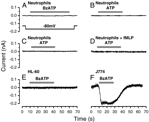
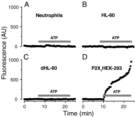
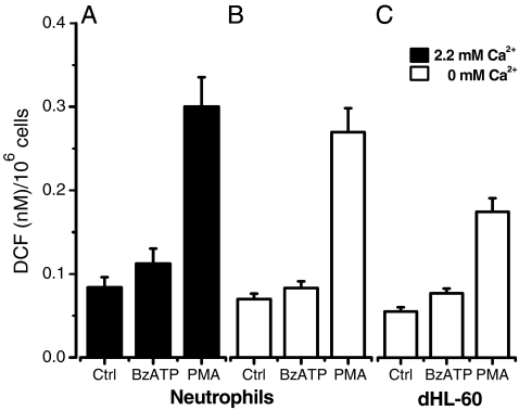
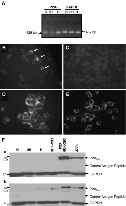
It has been reported that in human neutrophils, external ATP activates plasma membrane purinergic P2X(7) receptors (P2X(7)R) to elicit Ca(2+) entry, production of reactive oxygen species (ROS), processing and release of pro-inflammatory cytokines, shedding of adhesion molecules and uptake of large molecules. However, the expression of P2X(7)R at the plasma membrane of neutrophils has also been questioned since these putative responses are not always reproduced. In this work, we used electrophysiological recordings to measure functional responses associated with the activation of membrane receptors, spectrofluorometric measurements of ROS production and ethidium bromide uptake to asses coupling of P2X(7)R activation to downstream effectors, immune-labelling of P2X(7)R using a fluorescein isothiocyanate-conjugated antibody to detect the receptors at the plasma membrane, RT-PCR to determine mRNA expression of P2X(7)R and Western blot to determine protein expression in neutrophils and HL-60 cells. None of these assays reported the presence of P2X(7)R in the plasma membrane of neutrophils and non-differentiated or differentiated HL-60 cells-a model cell for human neutrophils. We concluded that P2X(7)R are not present at plasma membrane of human neutrophils and that the putative physiological responses triggered by external ATP should be reconsidered.

Keywords: Human; Neutrophils; Protein; Purinergic receptors; Whole cell current.


Figures

Fig. 1
Fig. 1
Purinergic stimulation does not induce development of currents in both neutrophils and HL-60 cells. A Representative (n = 4) current trace shows that no currents were induced after stimulating a human neutrophil with 100 μM BzATP. B No current was induced by stimulation of neutrophils with 250 μM ATP (n = 8). Currents were recorded using solutions containing 140 mM Na+. C Absence of ATP-activated current in a human neutrophil isolated in the presence of 2 U of apyrase (n = 6). D Pretreatment with 1 μM fMLP during 10 min at 37°C of human neutrophils isolated with apyrase did not result in currents after stimulation with 5 mM ATP (n = 3). E Representative current trace (n = 3) shows that in HL-60 cells, no currents were induced by stimulation with 100 μM BzATP. In this experiment, main cations were K+ (115 mM KCl) in the internal solution and TEA+ (140 mM TEACl) in the external solution. F Rapidly activating current recorded after stimulation of J774 cells with 100 µM BzATP (n = 9). Grey bars above each trace indicate duration of purinergic stimulation. All currents were recorded at −80 mV. The voltage protocol used is schematically represented in the inset of A
Fig. 2
Fig. 2
Lack of ATP-induced ethidium uptake in human neutrophils, undifferentiated HL-60 cells and differentiated HL-60 cells. AD Time course of the fluorescence signals resulting from ethidium uptake reported in arbitrary units (AU), from four different representative cells before and after stimulation. Purinergic stimulation did not induce ethidium uptake in human neutrophils (A) (n = 6), HL-60 cells (B) (n = 8) and differentiated HL-60 cells (C) (n = 8). HEK-293 cells transfected with mP2X7R (D) (n = 5), used here as a positive control, displayed large fluorescence increase, indicating ethidium uptake upon stimulation with ATP. Grey bars indicate stimulation with 5 mM ATP
Fig. 3
Fig. 3
BzATP did not induce ROS production in human neutrophils and differentiated HL-60 cells. Cells (106/ml) loaded with DCFH2-DA were treated with vehicle (control), 100 μM BzATP or 10 µM PMA in solutions containing either 2.2 mM (A) (n = 5) or 0 mM (B) (n = 11) extracellular Ca2+. C Lack of ROS production in differentiated HL-60 cells stimulated with 100 μM BzATP in the absence of extracellular Ca2+ (n = 7). As expected, treatment with PMA (10 µM) induced a large increase in fluorescence signal in all cells
Fig. 4
Fig. 4
Lack of P2X7R protein expression in neutrophils and HL-60 cells. A Detection of mRNA in human neutrophils (lane N), differentiated HL-60 cells (lane dH) and HL-60 cells (lane H). GAPDH was used as a positive control. The expected length of the PCR products obtained in these reactions is indicated next to the arrows. B, C P2X7R was not detected by antibody P2X7-ext at the surface of human neutrophils (B) (representative of n = 3) or of HL-60 cells (C) (n = 2). White arrows in B indicate three cells that are probably contamination and were labelled by the antibody. D, E Representative pictures of mP2X7-transfected HEK-293 (n = 4) and J774 (n = 2) cells used as positive controls for surface labelling of P2X7R. All cells were incubated with anti P2X7-FITC antibody (dilution 1:100) during 1 h as described in “Materials and methods”. F Western blots to determine P2X7R protein in total lysates of human neutrophils (lane N), differentiated HL-60 cells (lane dH), non-differentiated HL-60 cells (lane H), non-transfected HEK-293 cells (lane HEK-293), mP2X7R-transfected HEK-293 cells (lane P2X7HEK-293) and J774 macrophages (lane J774). Neither the P2X7-int antibody nor the P2X7ext antibody detected P2X7R protein in human neutrophils (n = 4), in non-differentiated (n = 4) or differentiated HL-60 cells (n = 4) or in non-transfected HEK-293 (n = 3). In contrast, in HEK-293 transfected with mP2X7R (n = 3) and J774 (n = 3) cells, the mP2X7R protein was readily detected. GAPDH served as load marker

Similar articles

Cited by

References

    1. Adrian K, Bernhard MK, Breitinger HG, Ogilvie A. Expression of purinergic receptors (ionotropic P2X1-7 and metabotropic P2Y1-11) during myeloid differentiation of HL60 cells. Biochim Biophys Acta. 2000;1492:127–138. - PubMed
    1. Akbar GK, Mills DC, Kunapuli SP. Characterization of extracellular nucleotide-induced Mac-1 (alphaM beta2 integrin) surface expression on peripheral blood leukocytes. Biochem Biophys Res Commun. 1997;233:71–75. doi: 10.1006/bbrc.1997.6396. - DOI - PubMed
    1. Axtell RA, Sandborg RR, Smolen JE, Ward PA, Boxer LA. Exposure of human neutrophils to exogenous nucleotides causes elevation in intracellular calcium, transmembrane calcium fluxes, and an alteration of a cytosolic factor resulting in enhanced superoxide production in response to FMLP and arachidonic acid. Blood. 1990;75:1324–1332. - PubMed
    1. Bass DA, Parce JW, Dechatelet LR, Szejda P, Seeds MC, Thomas M. Flow cytometric studies of oxidative product formation by neutrophils: a graded response to membrane stimulation. J Immunol. 1983;4:1910–1917. - PubMed
    1. Bours MJL, Swennen ELR, Virgilio F, Cronstein BN, Dagnelie PC. Adenosine 5’-triphosphate and adenosine as endogenous signalling molecules in immunity and inflammation. Pharmacol Ther. 2006;112:358–404. doi: 10.1016/j.pharmthera.2005.04.013. - DOI - PubMed

LinkOut - more resources