. 2010 Sep;6(3):307-15.
doi: 10.1007/s11302-010-9181-z. Epub 2010 Apr 21.

The effects of P2X7 receptor antagonists on the formation and function of human osteoclasts in vitro

The effects of P2X7 receptor antagonists on the formation and function of human osteoclasts in vitro

Ankita Agrawal et al. Purinergic Signal. 2010 Sep.

Abstract

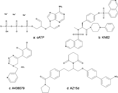
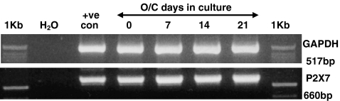
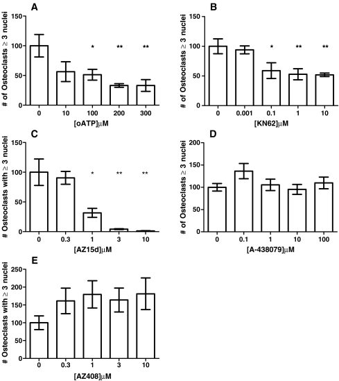
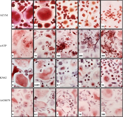
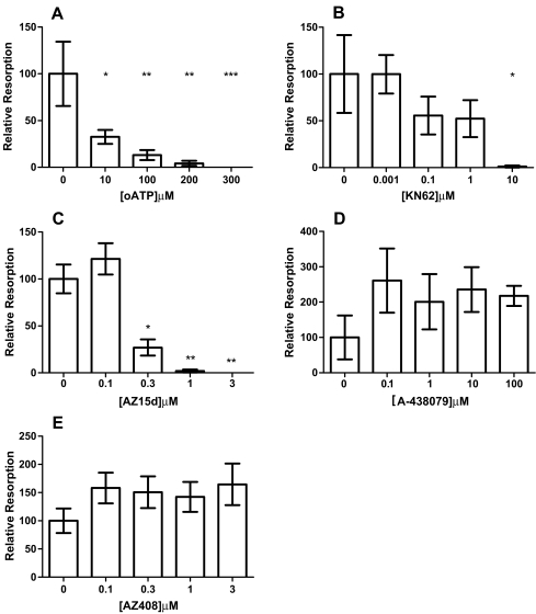
The P2X7 receptor (P2X7R) has been implicated in the process of multinucleation and cell fusion. We have previously demonstrated that blockade of P2X7Rs on osteoclast precursors using a blocking antibody inhibited multinucleated osteoclast formation in vitro, but that P2X7R KO mice maintain the ability to form multinucleated osteoclasts. This apparent contradiction of the role the P2X7R plays in multinucleation has prompted us to examine the effect of the most commonly used and recently available P2X7R antagonists on osteoclast formation and function. When added to recombinant RANKL and M-CSF human blood monocytes cultures, all but one compound, decreased the formation and function of multinucleated TRAP-positive osteoclasts in a concentration-dependent manner. These data provide further evidence for the role of the P2X7R in the formation of functional human multinucleated osteoclasts and highlight the importance of selection of antagonists for use in long-term experiments.

Keywords: ATP; Formation; Fusion; Osteoclast; P2 receptor; P2X7; Resorption.


Figures

Fig. 1
Fig. 1
Chemical structure of P2X7R antagonists. a oATP, b KN62, c A-438079 and d AZ15d
Fig. 2
Fig. 2
P2X7R mRNA is expressed during osteoclastogenesis in vitro. Osteoclasts were generated from monocytes cultured in the presence of recombinant M-CSF and RANKL. RNA was isolated from these cells after 0, 7, 14 and 21 days in culture. cDNA was generated and subsequently used as a template for PCR with primers designed specifically to the P2X7R. Resulting PCR products were separated by agarose gel electrophoresis and visualised under UV transillumination. +ve con = positive control, 1 Kb = 1 Kb molecular weight marker
Fig. 3
Fig. 3
Effect of P2X7R antagonists on human osteoclast formation. Osteoclasts were generated from human blood monocytes cultured on 6-mm coverslips in recombinant M-CSF and RANKL-supplemented medium. Antagonists were introduced throughout the culture by inclusion in the medium at each medium change at the concentrations shown on the x axis. Vehicle was complete α-MEM plus recombinant RANKL (30 ng/ml) and M-CSF (25 ng/ml) and 0.1% DMSO for KN62, AZ15d and AZ408. The number of osteoclasts was determined by counting all TRAP-positive cells with three or more nuclei. Data show means ± SEM, *p < 0.05, **p < 0.01 ***p < 0.0001 Graph representative of four repeat experiments, n = 7. a oATP, b KN62, c AZ15d, d A-438079 and e AZ408
Fig. 4
Fig. 4
P2X7R antagonists inhibit human osteoclast formation but are not toxic. Osteoclasts were generated from human blood monocytes cultured on 6-mm coverslips in recombinant M-CSF and RANKL-supplemented medium. Antagonists were introduced throughout the culture by inclusion in the medium at each medium change at the concentrations shown. Vehicle was complete α-MEM plus recombinant RANKL (30 ng/ml) and M-CSF (25 ng/ml) and 0.1% DMSO for KN62, and AZ15d. The coverslips were fixed in acetone and stained for tartrate-resistant acid phosphatase (TRAP) using a commercially available kit used according to the manufacturer’s protocol or fixed in formalin and stained as previously described [25]. Typical fields of view of cells following treatment in μM as indicated. Scale bar 30 μ
Fig. 5
Fig. 5
Effect of P2X7R antagonists on human osteoclast resorption. Osteoclasts were grown on dentine discs from blood monocytes in the presence of recombinant M-CSF and RANKL. Antagonists were introduced throughout the culture by inclusion in the medium at each medium change at the concentrations shown on the x axis. Vehicle was complete α-MEM plus recombinant RANKL (30 ng/ml) and M-CSF (25 ng/ml) and 0.1% DMSO for KN62, AZ15d and AZ408. After 3 weeks in culture, discs were fixed and stained with toluidine blue or TRAP. The area of resorption excavated by these cells was determined by point counting [26]. Data show means ± SEM, *p < 0.05, **p < 0.01 ***p < 0.0001. Graphs representative of four repeat experiments, n = 7. a oATP, b KN62, c AZ15d, d A-438079 and e AZ408

References

    1. Rassendren F, Buell GN, Virginio C, Collo G, North RA, Surprenant A. The permeabilizing ATP receptor, P2X7. Cloning and expression of a human cDNA. J Biol Chem. 1997;272:5482–5486. - PubMed
    1. Chessell IP, Michel AD, Humphrey PP. Effects of antagonists at the human recombinant P2X7 receptor. Br J Pharmacol. 1998;124:1314–1320. doi: 10.1038/sj.bjp.0701958. - DOI - PMC - PubMed
    1. Virgilio F. The P2Z purinoceptor: an intriguing role in immunity, inflammation and cell death. Immunol Today. 1995;16:524–528. doi: 10.1016/0167-5699(95)80045-X. - DOI - PubMed
    1. Surprenant A, Rassendren F, Kawashima E, North RA, Buell G. The cytolytic P2Z receptor for extracellular ATP identified as a P2X receptor (P2X7) Science. 1996;272:735–738. doi: 10.1126/science.272.5262.735. - DOI - PubMed
    1. Ferrari D, Chiozzi P, Falzoni S, Dal Susino M, Melchiorri L, Baricordi OR, Virgilio F. Extracellular ATP triggers IL-1 beta release by activating the purinergic P2Z receptor of human macrophages. J Immunol. 1997;159:1451–1458. - PubMed