

Intraabdominal Intravascular Papillary Endothelial Hyperplasia (Masson's Tumor): A Rare and Novel Cause of Gastrointestinal Bleeding
- PMID: 21103239
- PMCID: PMC2988909
- DOI: 10.1159/000294148
Intraabdominal Intravascular Papillary Endothelial Hyperplasia (Masson's Tumor): A Rare and Novel Cause of Gastrointestinal Bleeding
Abstract
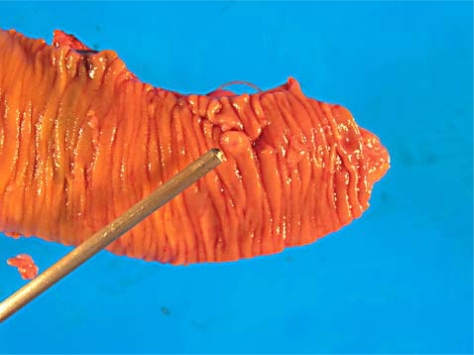
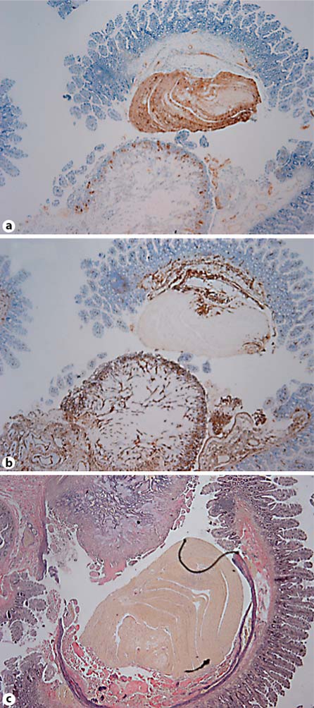
Intravascular papillary endothelial hyperplasia (IPEH), or Masson's tumor, a rare benign vascular lesion, occurs mainly in the head, neck, and hands in the human population. Aberrant tumor locations have been rarely reported. We present a case of a patient with chronic abdominal pain and melena of variable severity due to a Masson's tumor, with no apparent Masson's tumor-associated comorbidities, along with a comprehensive review of the literature. Using PubMed, a search engine provided by the U.S. National Library of Medicine and the National Institutes of Health, we searched for all reports of Masson's tumor limited within the abdominal cavity. Furthermore, keywords such as 'intravascular papillary endothelial hyperplasia', 'renal', 'gastrointestinal', 'hepatic' and 'intraabdominal' were used to facilitate the search. We thus found fourteen cases of intraabdominal Masson's tumors published. Six (42.9%) of these were located in the renal vein, 4 (28.6%) were reported in the gastrointestinal tract, 1 (7.1%) in the adrenal gland, 1 (7.1%) in the liver, and 1 (7.1%) instance with multiple lesion sites including the renal hilum and retroperitoneum. Among these patients, 9 (64.3%) were female and 5 (35.7%) male, with a mean age of 38.9 years (7-69). IPEH is a reactive process, having three subtypes, all involving the proliferation of epithelial cells around a thrombus in the setting of venous stasis. In its pure form, the organized thrombus is solely localized within the vascular lumen. Mixed-form IPEH is formed in preexisting vascular lesions (such as arteriovenous malformation, hemangioma, pyogenic granuloma, etc.). The rarest form is the extravascular variety, which arises in hematomas often from recent trauma to the area. In its pure form, IPEH has a zero recurrence rate when an R0 resection is performed; all mixed and extravascular forms show the highest recurrence rates. The exact histogenesis of these epithelial cells remains a mystery and multiple theories have been offered. Although difficult to distinguish from malignant angiosarcomas solely on presentation and radiologic work-up, Masson's tumors occur more frequently in women, demonstrate no local invasion, do not metastasize, are commonly located intravascularly, and are associated with a significantly more favorable prognosis than angiosarcomas. Only four Masson's tumors have been reported in the gastrointestinal tract, two of these cases were related to microvascular thrombosis secondary to paroxysmal nocturnal hemoglobinuria and two were formed secondary to arteriovenous malformations. Our case lacked solitary evidence of either of these comorbidities. An incidental finding of an enlarged ovary, which was removed during our exploratory laparoscopy, plus strong demographic statistics that suggest women have an increased prevalence of this lesion may help support a hormonal theory of pathogenesis.
Figures

References
-
- Hashimoto H, Daimaru Y, Enjoji M. Intravascular papillary endothelial hyperplasia: a clinicopathologic study of 91 cases. Am J Dermatopathol. 1938;5:539–546. - PubMed
-
- Clearkin KP, Enzinger FM. Intravascular papillary endothelial hyperplasia. Arch Pathol Lab Med. 1976;100:441–444. - PubMed
-
- Salyer WR, Salyer DC. Intravascular angiomatosis: Development and distinction from angiosarcoma. Cancer. 1975;36:995–1001. - PubMed
-
- Garber BB, Prestipino AJ, Pollack HM, Levine SR, Whitmore KE. Masson's tumor of the kidney: a new renal lesion. J Urol. 1990;143:344–346. - PubMed
-
- Steffe CH, Iskandar SS. Intravascular papillary endothelial hyperplasia in a thrombosed renal allograft vein. Hum Pathol. 1996;27:986–999. - PubMed
Publication types
LinkOut - more resources
Full Text Sources
Molecular Biology Databases
