

The society for gastrointestinal intervention. Are we, as an organization of disparate disciplines, cooperative or competitive?
- PMID: 21103287
- PMCID: PMC2989558
- DOI: 10.5009/gnl.2010.4.S1.S1
The society for gastrointestinal intervention. Are we, as an organization of disparate disciplines, cooperative or competitive?
Abstract
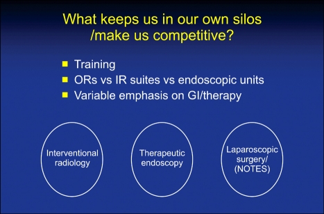
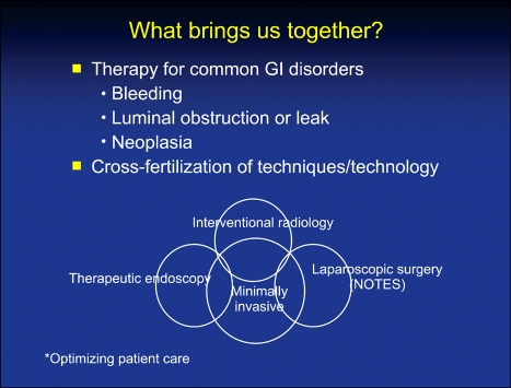
This is the Fourth Annual Meeting of the Society for Gastrointestinal Intervention, a multi-disciplinary group of practitioners committed to a minimally invasive approach to both the diagnosis and treatment of digestive disorders. The key concepts are minimally invasive and multi-disciplinary which can be construed as practicing in parallel with occasional lines of procedural and clinical interaction or inter-disciplinary in which patients are acutely cared for by a team, with treatments tailored to the patient and not the discipline that touches the patient first. In reality, many of us exist in both worlds. Most universities and large clinics are structured in departments along traditional training lines. As such, Interventional Radiology is housed in the Radiology Department, Laparoscopic Surgery (and potentially NOTES), as a component of the General Surgery Division, and Therapeutic Endoscopy usually resides within a gastroenterology structural framework. These divisions have historically been kept separate by multiple forces: salaries and budgets usually reside in a larger division. As a group, the amount of practice devoted to GI disorders is variable (for instance, minimally invasive surgeons may approach the adrenal glands or lung lesions in some institutions and interventional radiologists often sample tissue in multiple areas outside the GI tract, and by virtue of access to the vascular tree, can stent, embolize, or TPA almost any area of the body), as well as inherent differences in our individual abilities to access organs. I have already mentioned that angiographic capabilities allow the interventional radiologist access to virtually every GI organ and those capabilities allow therapeutic options for bleeding, tumor embolization, stenting of stenotic lesions, and formation of intravascular shunts. As such, there is very limited interdisciplinary competition here although capsule endoscopy as well as double and single balloon enteroscopy have improved the endoscopist's diagnostic and potential therapeutic reach. However, many of these diagnostic triumphs for obscure or massive GI bleed are simply to tattoo lesions that require surgical removal by laparoscopic or traditional surgery. Cooperation. However, there are potential competitive areas in the treatment of GI vascular lesions also. Whereas endoscopic band ligation has supplanted EVS, splenic devascularization, and most shunting procedures for patients with esophageal varices, endoscopic techniques have had less long-term success with glue injection for gastric varices. Multiple randomized, prospective trials have suggested therapeutic primacy of TIPS with embolization of recalcitrant vessels as an option or back-up. Despite this, therapeutic endoscopists have learned valuable lesions from our IR colleagues and studies are underway using endoscopically injected coils in addition to cyanoacrylate in an attempt to improve acute and long-term bleeding control. Nor is there any major competition in the treatment of primary or metastatic liver tumors by chemoembolization, RF current, or other thermal modalities, although selected patients with single lesions or multiple lesions isolated to a single lobe may be better handled surgically if there is curative intent. Finally, there is little IR, and progressively less, surgical competition for the treatment of high-grade dysplasia or superficial malignancies in the setting of Barrett's esophagus which are adequately treated in most patients by mucosectomy, RF ablation, or cryotherapy but require direct mucosal visualization to direct this therapy. The same has proven true for many years for colorectal polyps, superficial gastric cancers, and ampullary adenomas that had historically all been treated with major surgical resections. Still, there are many patients with advanced lesions who are good operative candidates who should be approached with conventional or minimally invasive surgery with the intent of operative cure. Cooperative, not competitive. The potential for competition between disciplines comes in mundane situations and clinical settings that have historically been "owned" by a single discipline. On the one hand, placement of PEGS and PEJs, initially done endoscopically, can be done with equal facility and occasional failure, by endoscopists and interventional radiologists, reserving failed attempts for minimally invasive surgery. What resources are utilized with these three methods? Are there advantages to defining the mucosa of the gut lumen in all, or even a subset of patients? By way of contrast, acute cholecystectomy tubes in high surgical risk patients have usually been the domain of the radiologist, although I described transcystic duct gallbladder decompression endoscopically 2½ decades ago. With the advent of new devices delivered under EUS control, the gallbladder will now be readily accessible endoscopically. What does this mean both for the acutely ill patient without a window to approach their gallbladder radiologically? Will this play a bit part and a cooperative technique to expand our therapeutic armamentarium or will it become competitive therapeutically not only for IR but for minimally invasive surgeons? The same may be said for EUS's ability to inject genes, caustics, or chemo-therapeutic agents into organs adjacent to the lumen. What is the role of TNFerade injection into unresectable pancreatic cancers and the role of absolute alcohol or Taxitol to treat cystic neoplasms of the pancreas? The real issue of competition or cooperation between the disciplines comes when treating patients with unresectable and obstructing GI neoplasms, from my perspective. The latter may occur almost anywhere in the GI tract but, of course, are more commonly noted proximally (esophagus, stomach, duodenum) and distally (left colon) as well as proximal and distal biliary obstructions. Recognizing that the occasional mid-small bowel and many proximal colon lesions are better handled with an endoscopic approach because of loss of vector force and difficulty pushing a catheter through large diameter, acutely angulated lumens, all others are fair game from my perspective. To my knowledge, although there are studies demonstrating the superiority of SEMS over open or laparoscopic bypass for malignant gastric outlet obstruction insofar as return of gut function, hospitalization time, and resource utilization, there are no studies demonstrating the superiority of one discipline or another in the placement of SEMS. Nor have cost data emerged suggesting the superiority of one technique over another from a cost standpoint. Unless or until we have such studies, this suggests to me that institutional interest and expertise should play a major role in how these unfortunate patients have continuity of their GI tract re-established. The situation is a bit more complex in pancreaticobiliary malignancy. There are 2 prospective randomized trials (level 1 evidence) that suggest that patients with proximal strictures (Bismuth II-IV) in conjunction with bile duct and gallbladder cancer, respectively, may be more successfully stented percutaneously and certainly it is easier to deliver brachytherapy or PDT under protocol to these patients who have indwelling external drains. In contrast, there are no data, positive or negative, to suggest that PTBD is a preferable treatment for distal biliary malignant obstruction, and in most parts of the world, the endoscopic approach has supplanted the percutaneous one just as metal stents have replaced plastic prostheses to preclude recurrent bouts of stent dysfunction and need for additional ERCP. The question posed at the beginning of this syllabus contribution: Are we competitive or cooperative? The answer is obviously both but, hopefully, our choice of treatment should depend less on who touches the patient first and more on skill sets within an institution and what is the best treatment for this particular individual. The importance of the SGI is technical and informational cross-fertilization. If your university or clinic will not allow blurring of training barriers to put therapeutic endoscopists, minimally invasive surgeons, and interventional radiologists together as a department or institute, you can nevertheless work together as a team in the best interest of your patients.
Keywords: Interventional radiology; Minimally invasive surgery; Therapeutic endoscopy.
Figures
Similar articles
-
American Gastroenterological Association Clinical Practice Update: Management of Pancreatic Necrosis.Gastroenterology. 2020 Jan;158(1):67-75.e1. doi: 10.1053/j.gastro.2019.07.064. Epub 2019 Aug 31. Gastroenterology. 2020. PMID: 31479658 Review.
-
The future of Cochrane Neonatal.Early Hum Dev. 2020 Nov;150:105191. doi: 10.1016/j.earlhumdev.2020.105191. Epub 2020 Sep 12. Early Hum Dev. 2020. PMID: 33036834
-
Endoscopic foregut surgery and interventions: The future is now. The state-of-the-art and my personal journey.World J Gastroenterol. 2019 Jan 7;25(1):1-41. doi: 10.3748/wjg.v25.i1.1. World J Gastroenterol. 2019. PMID: 30643356 Free PMC article. Review.
-
Port site metastases a year after initial laparoscopic cholecystectomy. Should the use of retrieval bags during laparoscopic cholecystectomy be the new gold standard?Pol Przegl Chir. 2021 May 31;93(6):61-65. doi: 10.5604/01.3001.0015.3280. Pol Przegl Chir. 2021. PMID: 36169533
-
AGA Institute Clinical Practice Update: Endoscopic Submucosal Dissection in the United States.Clin Gastroenterol Hepatol. 2019 Jan;17(1):16-25.e1. doi: 10.1016/j.cgh.2018.07.041. Epub 2018 Aug 2. Clin Gastroenterol Hepatol. 2019. PMID: 30077787 Review.
Cited by
-
Laparoscopic and endoscopic cooperative surgery for gastric tumors: Perspective for actual practice and oncological benefits.World J Gastrointest Oncol. 2018 Nov 15;10(11):381-397. doi: 10.4251/wjgo.v10.i11.381. World J Gastrointest Oncol. 2018. PMID: 30487950 Free PMC article. Review.
-
Laparoscopic implementation of the Altemeier procedure for recurrent rectal prolapse. Technical note.Int J Surg Case Rep. 2014;5(7):347-9. doi: 10.1016/j.ijscr.2014.04.011. Epub 2014 Apr 15. Int J Surg Case Rep. 2014. PMID: 24846791 Free PMC article.
-
The Business Engineering Surgical Technologies (BEST) teaching method: incubating talents for surgical innovation.Surg Endosc. 2015 Jan;29(1):48-54. doi: 10.1007/s00464-014-3652-1. Epub 2014 Jul 4. Surg Endosc. 2015. PMID: 24993171 Review.
-
Percutaneous transhepatic or endoscopic ultrasound-guided biliary drainage in malignant distal bile duct obstruction using a self-expanding metal stent: Study protocol for a prospective European multicenter trial (PUMa trial).PLoS One. 2022 Oct 27;17(10):e0275029. doi: 10.1371/journal.pone.0275029. eCollection 2022. PLoS One. 2022. PMID: 36302047 Free PMC article.
References
-
- Martinez Rodrigo J, Marti-Bonmati L, Segarra Medrano A, et al. Certification guidelines of the Spanish Society of Diagnostic Radiology (SERAM) and the Spanish Society of Vascular and Interventional Radiology (SERVEI) concerning requirements and equipment in vascular and interventional radiology. Radiologia. 2007;49:381–387. - PubMed
-
- Desser TS. Simulation-based training: the next revolution in radiology education? J Am Coll Radiol. 2007;4:816–824. - PubMed
-
- Hamad GG, Curet M. Minimally invasive surgery. Am J Surg. 2010;199:263–265. - PubMed
-
- Stefanidis D, Hope WW, Korndorffer JR, Jr, Markley S, Scott DJ. Initial laparoscopic basic skills training shortens the learning curve of laparoscopic suturing and is cost-effective. J Am Coll Surg. 2010;210:436–440. - PubMed
-
- Bittner JG, 4th, Coverdill JE, Imam T, Deladisma AM, Edwards MA, Mellinger JD. Do increased training requirements in gastrointestinal endoscopy and advanced laparoscopy necessitate a paradigm shift? A survey of program directors in surgery. J Surg Educ. 2008;65:418–430. - PubMed
LinkOut - more resources
Full Text Sources
Other Literature Sources
Research Materials
