. 2010 Nov 16;5(11):e15503.
doi: 10.1371/journal.pone.0015503.

Tyr(682) in the intracellular domain of APP regulates amyloidogenic APP processing in vivo

Affiliations

Tyr(682) in the intracellular domain of APP regulates amyloidogenic APP processing in vivo

Alessia P M Barbagallo et al. PLoS One. .

Abstract

Background: The pathogenesis of Alzheimer's disease is attributed to misfolding of Amyloid-β (Aβ) peptides. Aβ is generated during amyloidogenic processing of Aβ-precursor protein (APP). Another characteristic of the AD brain is increased phosphorylation of APP amino acid Tyr(682). Tyr(682) is part of the Y(682)ENPTY(687) motif, a docking site for interaction with cytosolic proteins that regulate APP metabolism and signaling. For example, normal Aβ generation and secretion are dependent upon Tyr(682) in vitro. However, physiological functions of Tyr(682) are unknown.

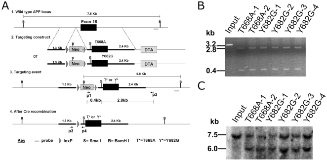
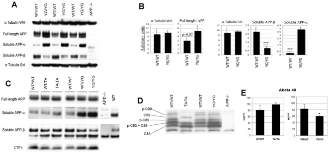
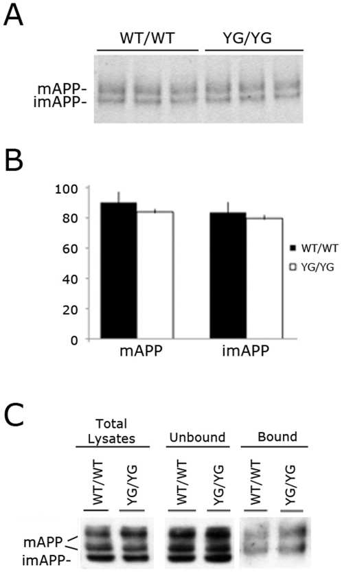
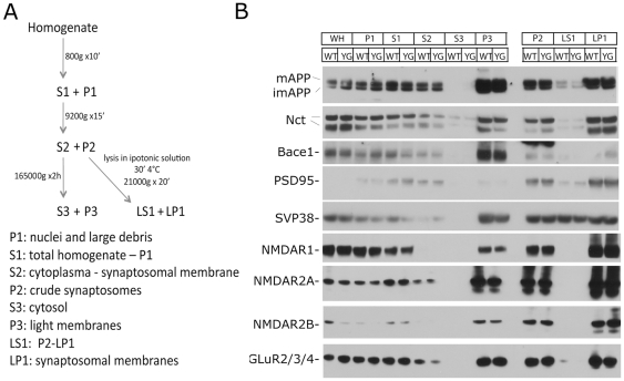
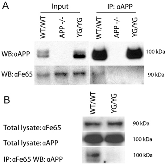
Methodology/principal findings: To this end, we have generated an APP Y682G knock-in (KI) mouse to help dissect the role of APP Tyr(682) in vivo. We have analyzed proteolytic products from both the amyloidogenic and non-amyloidogenic processing of APP and measure a profound shift towards non-amyloidogenic processing in APP KI mice. In addition, we demonstrate the essential nature of amino acid Tyr(682) for the APP/Fe65 interaction in vivo.

Conclusions/significance: Together, these observations point to an essential role of APP intracellular domain for normal APP processing and function in vivo, and provide rationale for further studies into physiological functions associated with this important phosphorylation site.


Conflict of interest statement

Competing Interests: The authors have declared that no competing interests exist.

Figures

Figure 1
Figure 1. Generation of APPYG mice.
A, Schematic representation of the construct injected in 129 ES cells, showing site of APP T668A and Y682G mutation on last Exon 16, primer sites, site of Southern Blot probe, LoxP, pgk-dta and pgk-Neo sites. The bottom graphics depict the construct with and without the pgk-Neo cassette that has been removed by means of Cre recombinase. B, The right arm (p1–p2) PCR analysis of six positively targeted ES clones. A 3.2 Kb PCR product digested by a novel restriction site Sma I produce 0.4 Kb and 2.8 Kb fragment. C, Southern Blot showing a shift from the 7.5 Kb of the wild type genome to the 6.0 Kb band of two T668A and four Y682G positively knock in ES clones for the homologous recombination of the mutated allele, due to the insertion of a new BamHI site.
Figure 2
Figure 2. Altered APP proteolytic processing in APPYG/YG mouse brain.
APP−/− brain was used as a negative control in each experiment. A, Immunoblot analysis comparing full length APP (WH  =  whole homogenates), sAPP-α, sAPP-b and tubulin levels between APPwt/wt APPYG/YG mice (n = 3). B, Quantitative analysis of panel A normalized to tubulin (***  =  p<0.01). C, WB analysis of APPwt/wt, APPwt/TA, APPTA/TA, APPwt/YG and APPYG/YG brain lysate showing full length APP, sAPP-α and sAPP-β. D, WB analysis of APP CTF's using a tris-tricine gel, and comparing APPwt/wt with APPTA/TA and APPYG/YG. Five specific species representing C83, C89, C99 and their respective phosphorylated forms can be identified, noting that p-C83 and C89 overlap. Bands at the very top and bottom are non-specific. E, Aβ4ELISA comparing APPwt/wt with APPTA/TA (n = 3) and APPwt/wt with APPYG/YG (n = 4). APP−/− mice were also used to validate specificity of the assay (data not shown).
Figure 3
Figure 3. Normal expression of the mutant APP allele.
A, The genetic manipulation of the APP gene locus in APPYG/YG mice does not affect transcription/splicing of APP since mRNA APP levels are comparable to those transcribed in age-matched APPWT/WT mice (analogous data were obtained using Beta Actin as housekeeping gene). B, APP and APP-derived CTFs are processed by the lysosomes. Inhibition of lysosomal activity by chloroquine (Chl.) results in accumulation of APP and APP derived fragments. The asterisks indicate APP-CTFs that are derived by cleavages in the ectodomain NH2-terminal to the site processed by BACE1. The effectiveness of Chl. in inhibiting lysosomal degradation (inhibition occurs at 50 µM concentration but not at 5 mM) is confirmed by the accumulation on LC3II . The antibody against LC3 is from Cell Signaling.
Figure 4
Figure 4. Maturation and membrane levels of APP are unaffected by the YG mutation.
A, Immunoblot analysis comparing imAPP and mAPP between APPwt/wt and APPYG/YG mice (n = 3). B, Quantitative analysis of panel A shows no differences in mAPP and imAPP levels between the two genotypes. C, Biotynilation experiment in APPwt/wt and APPYG/YG MDFs shows comparable levels of im and mAPP, as well as cell membrane levels of mAPP.
Figure 5
Figure 5. Alterations in quadriceps muscle from two APPYG/YG mice.
A, normal striated muscle. B, muscle fibers undergoing regeneration.
Figure 6
Figure 6. Coronal sections comparing brain anatomy of APPwt/wt with APPYG/YG mice.
A–E coronal sections of APPwt/wt mouse, F–J coronal sections of APPYG/YGmouse. A, F - Olfactory bulbs. B, G- Cerebral cortex. C, H- Thalamus. D, I- Midbrain. E, J- Medulla. Ob-olfactory bulb, r-retina, cc- cerebral cortex, s- striatum, hi- hippocampus, th- thalamus, mb- midbrain, ce- cerebellum.
Figure 7
Figure 7. The YG mutation does not alter synaptic proteins.
A, Schematic representation of synaptosomal preparation. B, Synaptic proteins distribution is similar in APPwt/wt and APPYG/YG brains.
Figure 8
Figure 8. Loss of APP/Fe65 interaction in APPYG/YG mouse brain.
A, IP of APP from a membrane enriched brain fraction using anti-APP antibody. WB analysis for Fe65 shows an absence of detectable signal in both APP−/− and APPYG/YG compared to APPwt/wt. B, IP of Fe65 from a membrane enriched brain fraction using anti-Fe65 antibody (I12). WB analysis for APP shows an absence of detectable signal in APPYG/YG mutant mice compared to APPwt/wt.

References

    1. Hardy JA, Higgins GA. Alzheimer's disease: the amyloid cascade hypothesis. Science. 1992;256:184–185. - PubMed
    1. Selkoe DJ. The genetics and molecular pathology of Alzheimer's disease: roles of amyloid and the presenilins. Neurol Clin. 2000;18:903–922. - PubMed
    1. Passer B, Pellegrini L, Russo C, Siegel RM, Lenardo MJ, et al. Generation of an apoptotic intracellular peptide by gamma-secretase cleavage of Alzheimer's amyloid beta protein precursor. J Alzheimers Dis. 2000;2:289–301. - PubMed
    1. Cao X, Sudhof TC. A transcriptionally [correction of transcriptively] active complex of APP with Fe65 and histone acetyltransferase Tip60. Science. 2001;293:115–120. - PubMed
    1. Stokin GB, Lillo C, Falzone TL, Brusch RG, Rockenstein E, et al. Axonopathy and transport deficits early in the pathogenesis of Alzheimer's disease. Science. 2005;307:1282–1288. - PubMed

Publication types

MeSH terms

LinkOut - more resources