

Intraspecies prion transmission results in selection of sheep scrapie strains
- PMID: 21103326
- PMCID: PMC2982847
- DOI: 10.1371/journal.pone.0015450
Intraspecies prion transmission results in selection of sheep scrapie strains
Abstract
Background: Sheep scrapie is caused by multiple prion strains, which have been classified on the basis of their biological characteristics in inbred mice. The heterogeneity of natural scrapie prions in individual sheep and in sheep flocks has not been clearly defined.
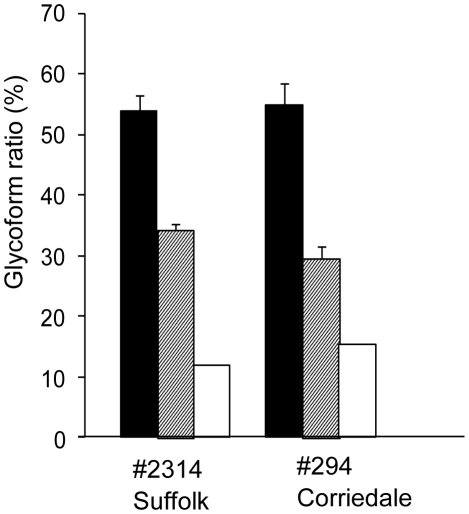
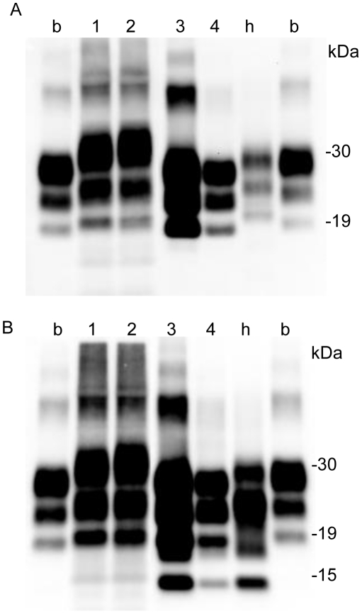
Methodology/principal findings: In this study, we intravenously injected 2 sheep (Suffolk and Corriedale) with material from a natural case of sheep scrapie (Suffolk breed). These 3 sheep had identical prion protein (PrP) genotypes. The protease-resistant core of PrP (PrPres) in the experimental Suffolk sheep was similar to that in the original Suffolk sheep. In contrast, PrPres in the Corriedale sheep differed from the original PrPres but resembled the unusual scrapie isolate, CH1641. This unusual PrPres was not detected in the original sheep. The PrPres distributions in the brain and peripheral tissues differed between the 2 breeds of challenged sheep. A transmission study in wild-type and TgBoPrP mice, which overexpressing bovine PrP, led to the selection of different prion strains. The pathological features of prion diseases are thought to depend on the dominantly propagated strain.
Conclusions/significance: Our results indicate that prion strain selection occurs after both inter- and intraspecies transmission. The unusual scrapie prion was a hidden or an unexpressed component in typical sheep scrapie.
Conflict of interest statement
Figures
Similar articles
-
Interspecies transmission to bovinized transgenic mice uncovers new features of a CH1641-like scrapie isolate.Vet Res. 2018 Nov 28;49(1):116. doi: 10.1186/s13567-018-0611-1. Vet Res. 2018. PMID: 30486902 Free PMC article.
-
Change in the molecular properties of CH1641 prions after transmission to wild-type mice: Evidence for a single strain.Neuropathol Appl Neurobiol. 2024 Feb;50(1):e12963. doi: 10.1111/nan.12963. Neuropathol Appl Neurobiol. 2024. PMID: 38353056
-
A C-terminal protease-resistant prion fragment distinguishes ovine "CH1641-like" scrapie from bovine classical and L-Type BSE in ovine transgenic mice.PLoS Pathog. 2008 Aug 29;4(8):e1000137. doi: 10.1371/journal.ppat.1000137. PLoS Pathog. 2008. PMID: 18769714 Free PMC article.
-
Genetic and infectious prion diseases.Arch Neurol. 1993 Nov;50(11):1129-53. doi: 10.1001/archneur.1993.00540110011002. Arch Neurol. 1993. PMID: 8105771 Review.
-
Prion encephalopathies of animals and humans.Dev Biol Stand. 1993;80:31-44. Dev Biol Stand. 1993. PMID: 8270114 Review.
Cited by
-
Distinct proteinase K-resistant prion protein fragment in goats with no signs of disease in a classical scrapie outbreak.J Clin Microbiol. 2011 Jun;49(6):2109-15. doi: 10.1128/JCM.02033-10. Epub 2011 Mar 30. J Clin Microbiol. 2011. PMID: 21450953 Free PMC article.
-
Selective propagation of mouse-passaged scrapie prions with long incubation period from a mixed prion population using GT1-7 cells.PLoS One. 2017 Jun 21;12(6):e0179317. doi: 10.1371/journal.pone.0179317. eCollection 2017. PLoS One. 2017. PMID: 28636656 Free PMC article.
-
Sheep prions with molecular properties intermediate between classical scrapie, BSE and CH1641-scrapie.Prion. 2014;8(4):296-305. doi: 10.4161/19336896.2014.983396. Prion. 2014. PMID: 25522672 Free PMC article.
-
An overview of animal prion diseases.Virol J. 2011 Nov 1;8:493. doi: 10.1186/1743-422X-8-493. Virol J. 2011. PMID: 22044871 Free PMC article. Review.
-
Ability of wild type mouse bioassay to detect bovine spongiform encephalopathy (BSE) in the presence of excess scrapie.Acta Neuropathol Commun. 2015 Apr 3;3:21. doi: 10.1186/s40478-015-0194-2. Acta Neuropathol Commun. 2015. PMID: 25853789 Free PMC article.
References
-
- Prusiner SB. Molecular biology of prion diseases. Science. 1991;252:1515–1522. - PubMed
-
- Caughey B, Raymond GJ. The scrapie-associated form of PrP is made from a cell surface precursor that is both protease- and phospholipase-sensitive. J Biol Chem. 1991;266:18217–18223. - PubMed
-
- Collinge J, Sidle KC, Meads J, Ironside J, Hill AF. Molecular analysis of prion strain variation and the aetiology of ‘new variant’ CJD. Nature. 1996;383:685–690. - PubMed
-
- Hayashi HK, Yokoyama T, Takata M, Iwamaru Y, Imamura M, et al. The N-terminal cleavage site of PrPSc from BSE differs from that of PrPSc from scrapie. Biochem Biophys Res Commun. 2005;328:1024–1027. - PubMed
Publication types
MeSH terms
Substances
LinkOut - more resources
Full Text Sources
Research Materials
