

Primary and malignant cholangiocytes undergo CD40 mediated Fas dependent apoptosis, but are insensitive to direct activation with exogenous Fas ligand
- PMID: 21103345
- PMCID: PMC2984448
- DOI: 10.1371/journal.pone.0014037
Primary and malignant cholangiocytes undergo CD40 mediated Fas dependent apoptosis, but are insensitive to direct activation with exogenous Fas ligand
Abstract
Introduction: Cholangiocarcinoma is a rare malignancy of the biliary tract, the incidence of which is rising, but the pathogenesis of which remains uncertain. No common genetic defects have been described but it is accepted that chronic inflammation is an important contributing factor. We have shown that primary human cholangiocyte and hepatocyte survival is tightly regulated via co-operative interactions between two tumour necrosis family (TNF) receptor family members; CD40 and Fas (CD95). Functional deficiency of CD154, the ligand for CD40, leads to a failure of clearance of biliary tract infections and a predisposition to cholangiocarcinoma implying a direct link between TNF receptor-mediated apoptosis and the development of cholangiocarcinoma.
Aims: To determine whether malignant cholangiocytes display defects in CD40 mediated apoptosis. By comparing CD40 and Fas-mediated apoptosis and intracellular signalling in primary human cholangiocytes and three cholangiocyte cell lines.
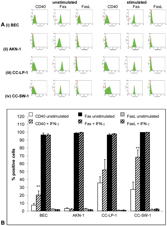
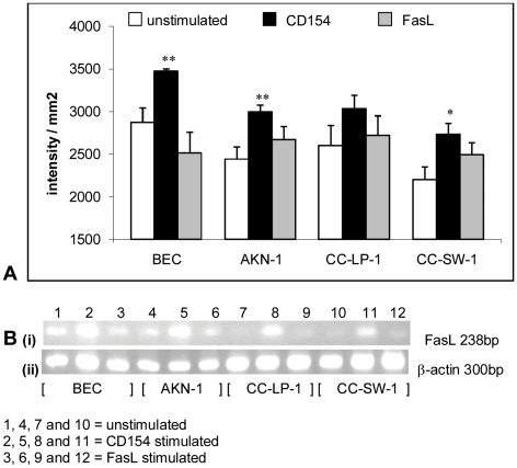
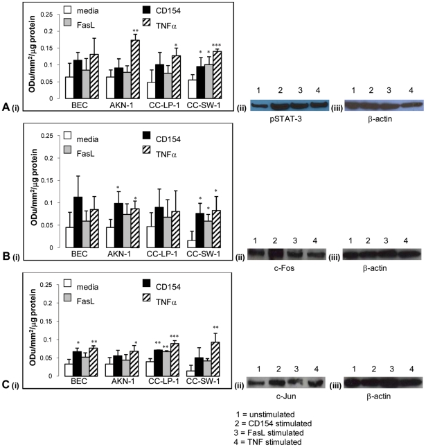
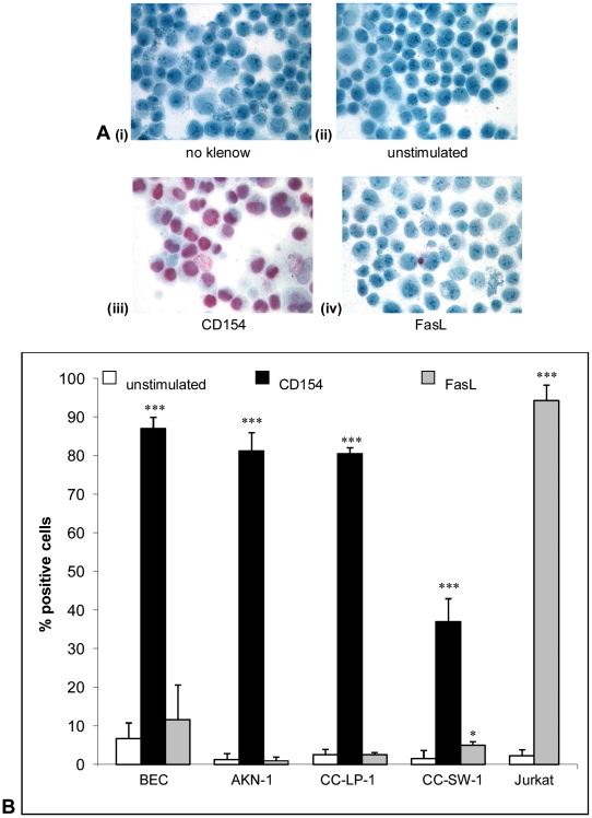
Results: Primary cholangiocytes and cholangiocyte cell lines were relatively insensitive to direct Fas-mediated killing with exogenous FasL when compared with Jurkat cells, which readily underwent Fas-mediated apoptosis, but were extremely sensitive to CD154 stimulation. The sensitivity of cells to CD40 activation was similar in magnitude in both primary and malignant cells and was STAT-3 and AP-1 dependent in both.
Conclusions: 1) Both primary and malignant cholangiocytes are relatively resistant to Fas-mediated killing but show exquisite sensitivity to CD154, suggesting that the CD40 pathway is intact and fully functional in both primary and malignant cholangiocytes 2) The relative insensitivity of cholangiocytes to Fas activation demonstrates the importance of CD40 augmentation of Fas dependent death in these cells. Agonistic therapies which target CD40 and associated intracellular signalling pathways may be effective in promoting apoptosis of malignant cholangiocytes.
Conflict of interest statement
Figures
References
-
- Itoh N, Yonehara S, Ishii A, Yonehara M, Mizushima S, et al. The polypeptide encoded by the cDNA for human cell surface antigen Fas can mediate apoptosis. Cell. 1991;66:233–243. - PubMed
-
- Suda T, Takahashi P, Goldstein P, Nagata S. Molecular cloning and expression of Fas ligand, a novel member of the tumour necrosis family. Cell. 1993;75:1169–1178. - PubMed
-
- French LE, Hahne M, Viarde I, Radlgrubber G, Zanone R, et al. Fas and Fas ligand in embryos and adult mice: ligand expression in several immune-privileged tissues and co expression in adult tissues characterized by apoptotic cell turnover. Journal of Cell Biology. 1996;133(2):335–343. - PMC - PubMed
-
- Bonfoco E, Stuart PM, Brunner T, Lin T, Griffith TS, et al. Inducible non-lymphoid expression of Fas ligand is responsible for superantigen-induced peripheral deletion of T cells. Immunity. 1998;9:711–720. - PubMed
-
- Harada K, Ozaki S, Gershwin ME, Nakanuma Y. Enhanced apoptosis relates to bile duct loss in primary biliary cirrhosis. Hepatology. 1997;26(6):1399–1405. - PubMed
Publication types
MeSH terms
Substances
Grants and funding
LinkOut - more resources
Full Text Sources
Other Literature Sources
Research Materials
Miscellaneous
