

Evidence for a transport-trap mode of Drosophila melanogaster gurken mRNA localization
- PMID: 21103393
- PMCID: PMC2980492
- DOI: 10.1371/journal.pone.0015448
Evidence for a transport-trap mode of Drosophila melanogaster gurken mRNA localization
Abstract
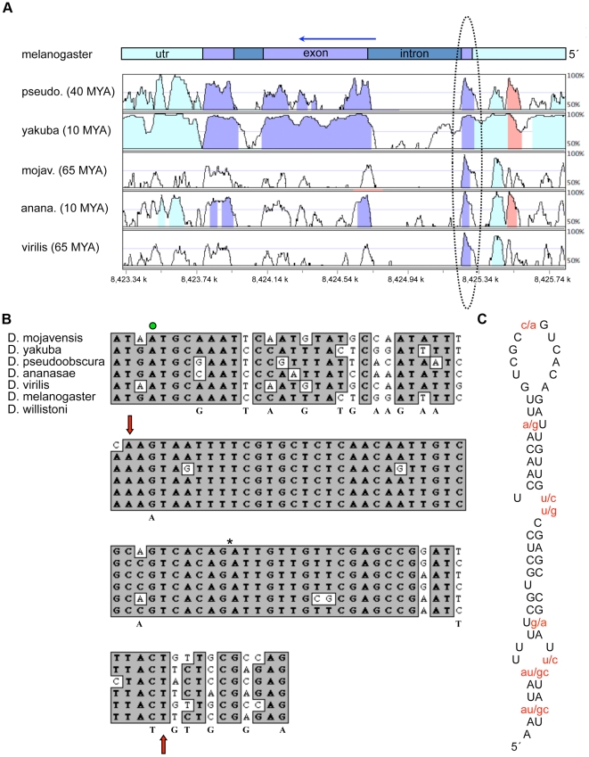
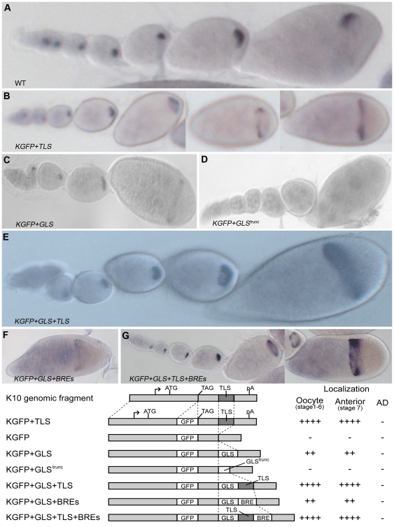
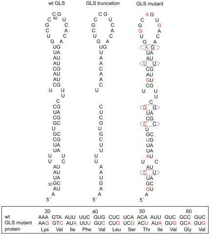
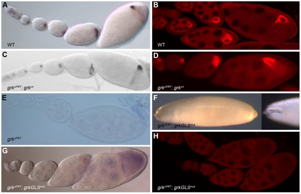
The Drosophila melanogaster gurken gene encodes a TGF alpha-like signaling molecule that is secreted from the oocyte during two distinct stages of oogenesis to define the coordinate axes of the follicle cell epithelium that surrounds the oocyte and its 15 anterior nurse cells. Because the gurken receptor is expressed throughout the epithelium, axial patterning requires region-specific secretion of Gurken protein, which in turn requires subcellular localization of gurken transcripts. The first stage of Gurken signaling induces anteroposterior pattern in the epithelium and requires the transport of gurken transcripts from nurse cells into the oocyte. The second stage of Gurken signaling induces dorsovental polarity in the epithelium and requires localization of gurken transcripts to the oocyte's anterodorsal corner. Previous studies, relying predominantly on real-time imaging of injected transcripts, indicated that anterodorsal localization involves transport of gurken transcripts to the oocyte's anterior cortex followed by transport to the anterodorsal corner, and anchoring. Such studies further indicated that a single RNA sequence element, the GLS, mediates both transport steps by facilitating association of gurken transcripts with a cytoplasmic dynein motor complex. Finally, it was proposed that the GLS somehow steers the motor complex toward that subset of microtubules that are nucleated around the oocyte nucleus, permitting directed transport to the anterodorsal corner. Here, we re-investigate the role of the GLS using a transgenic fly assay system that includes use of the endogenous gurken promoter and biological rescue as well as RNA localization assays. In contrast to previous reports, our studies indicate that the GLS is sufficient for anterior localization only. Our data support a model in which anterodorsal localization is brought about by repeated rounds of anterior transport, accompanied by specific trapping at the anterodorsal cortex. Our data further indicate that trapping at the anterodorsal corner requires at least one as-yet-unidentified gurken RLE.
Conflict of interest statement
Figures
Similar articles
-
The role of fs(1)K10 in the localization of the mRNA of the TGF alpha homolog gurken within the Drosophila oocyte.Mech Dev. 1995 Jun;51(2-3):183-92. doi: 10.1016/0925-4773(95)00363-0. Mech Dev. 1995. PMID: 7547466
-
Production of gurken in the nurse cells is sufficient for axis determination in the Drosophila oocyte.Development. 2005 May;132(10):2345-53. doi: 10.1242/dev.01820. Epub 2005 Apr 13. Development. 2005. PMID: 15829517
-
Drosophila gurken (TGFalpha) mRNA localizes as particles that move within the oocyte in two dynein-dependent steps.Dev Cell. 2003 Mar;4(3):307-19. doi: 10.1016/s1534-5807(03)00058-3. Dev Cell. 2003. PMID: 12636913
-
[Source of asymmetry in ontogeny: early polarization of the germline cyst and oocyte in Drosophila].Genetika. 2008 Sep;44(9):1157-71. Genetika. 2008. PMID: 18846812 Review. Russian.
-
The Role of Microtubule Motors in mRNA Localization and Patterning Within the Drosophila Oocyte.Results Probl Cell Differ. 2017;63:149-168. doi: 10.1007/978-3-319-60855-6_7. Results Probl Cell Differ. 2017. PMID: 28779317 Review.
Cited by
-
mRNA localization and translational control in Drosophila oogenesis.Cold Spring Harb Perspect Biol. 2012 Oct 1;4(10):a012294. doi: 10.1101/cshperspect.a012294. Cold Spring Harb Perspect Biol. 2012. PMID: 22865893 Free PMC article. Review.
-
mRNA localization in the Drosophila germline.RNA Biol. 2014;11(8):1010-8. doi: 10.4161/rna.36097. Epub 2014 Oct 31. RNA Biol. 2014. PMID: 25482896 Free PMC article. Review.
-
Control of cell migration through mRNA localization and local translation.Wiley Interdiscip Rev RNA. 2015 Jan-Feb;6(1):1-15. doi: 10.1002/wrna.1265. Epub 2014 Sep 28. Wiley Interdiscip Rev RNA. 2015. PMID: 25264217 Free PMC article. Review.
-
NineTeen Complex-subunit Salsa is required for efficient splicing of a subset of introns and dorsal-ventral patterning.RNA. 2020 Dec;26(12):1935-1956. doi: 10.1261/rna.077446.120. Epub 2020 Sep 22. RNA. 2020. PMID: 32963109 Free PMC article.
-
Finishing the egg.Genetics. 2024 Jan 3;226(1):iyad183. doi: 10.1093/genetics/iyad183. Genetics. 2024. PMID: 38000906 Free PMC article.
References
-
- Lecuyer E, Yoshida H, Parthasarathy N, Alm C, Babak T, et al. Global analysis of mRNA localization reveals a prominent role in organizing cellular architecture and function. Cell. 2007;131:174–187. - PubMed
-
- St Johnston D. Moving messages: the intracellular localization of mRNAs. Nat Rev Mol Cell Biol. 2005;6:363–375. - PubMed
-
- Kugler JM, Lasko P. Localization, anchoring and translational control of oskar, gurken, bicoid and nanos mRNA during Drosophila oogenesis. Fly (Austin) 2009;3:15–28. - PubMed
Publication types
MeSH terms
Substances
Grants and funding
LinkOut - more resources
Full Text Sources
Molecular Biology Databases
Miscellaneous
