. 2010 Oct;16(4):374-88.
doi: 10.5056/jnm.2010.16.4.374. Epub 2010 Oct 30.

Viewpoints on Acid-induced inflammatory mediators in esophageal mucosa

Affiliations

Viewpoints on Acid-induced inflammatory mediators in esophageal mucosa

Karen M Harnett et al. J Neurogastroenterol Motil. 2010 Oct.

Abstract

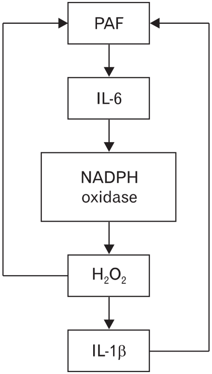
We have focused on understanding the onset of gastroesophageal reflux disease by examining the mucosal response to the presence of acid in the esophageal lumen. Upon exposure to HCl, inflammation of the esophagus begins with activation of the transient receptor potential channel vanilloid subfamily member-1 (TRPV1) in the mucosa, and production of IL-8, substance P (SP), calcitonin gene related peptide (CGRP) and platelet activating factor (PAF). Production of SP and CGRP, but not PAF, is abolished by the neural blocker tetrodotoxin suggesting that SP and CGRP are neurally released and that PAF arises from non neural pathways. Epithelial cells contain TRPV1 receptor mRNA and protein and respond to HCl and to the TRPV1 agonist capsaicin with production of PAF. PAF, SP and IL-8 act as chemokines, inducing migration of peripheral blood leukocytes. PAF and SP activate peripheral blood leukocytes inducing the production of H(2)O(2). In circular muscle, PAF causes production of IL-6, and IL-6 causes production of additional H(2)O(2), through activation of reduced nicotinamide adenine dinucleotide phosphate (NADPH) oxidases. Among these, NADPH oxidase 5 cDNA is significantly up-regulated by exposure to PAF; H(2)O(2) content of esophageal and lower esophageal sphincter circular muscle is elevated in human esophagitis, causing dysfunction of esophageal circular muscle contraction and reduction in esophageal sphincter tone. Thus esophageal keratinocytes, that constitute the first barrier to the refluxate, may also serve as the initiating cell type in esophageal inflammation, secreting inflammatory mediators and pro-inflammatory cytokines and affecting leukocyte recruitment and activity.

Keywords: Cytokines; Gastroesophageal reflux disease; Platelet activating factor; Substance P; TRPV1.


Conflict of interest statement

Conflicts of interest: None.

Figures

Figure 1
Figure 1
We produced an in vitro model of esophageal inflammation using an esophageal mucosa tube tied at both ends. The tube was created by removing the muscle layers from the esophagus by sharp dissection at the interface between submucosa and circular mucle. It has the squamous epithelium on the inside and the whole submucosa on the outside. HCl or Krebs buffer was injected inside the tube, which was then kept in oxygenated Krebs buffer at 36℃ for 3 hours (1 mL Krebs buffer/100 mg mucosa). After 3 hours the supernatant outside the tube was collected and analyzed or used to incubate circular muscle strips. Adapted from Cheng et al.
Figure 2
Figure 2
When in vitro mucosa is exposed to HCl for 3 hours, substance P levels increase 6-fold in the mucosa (A) and in the supernatant (B) when compared to control (pH 7.4) (*p < 0.05, ANOVA). The HCl-induced increase in substance P is abolished by the transient receptor potential channel vanilloid subfamily member-1 receptor antagonist 5'-iodoresinferratoxin (IRTX) and by the neural blocker tetrodotoxin (TTX) both in the mucosa and in the supernatant (**p < 0.05, ANOVA). Bars represent mean ± SEM of 3 experiments. Adapted from Cheng et al.
Figure 3
Figure 3
When in vitro mucosa is exposed to HCl for 3 hours, calcitonin gene related peptide (CGRP) levels increase 200-fold in the mucosa (A) and 10-fold in the supernatant (B) when compared to control (pH 7.4) (*p < 0.05, ANOVA). The HCl-induced increase in CGRP is abolished by the transient receptor potential channel vanilloid subfamily member-1 receptor antagonist 5'-iodoresinferratoxin (IRTX) and by the neural blocker tetrodotoxin (TTX) both in the mucosa and in the supernatant (**p < 0.05, ANOVA). Bars represent mean ± SEM of 3 experiments. Adapted from Cheng et al.
Figure 4
Figure 4
Histological sections of cat esophagus immunostained to detect (A, B) calcitonin gene related peptide (CGRP) or (C, D) Substance P. Immunoreactivity was detected with secondary antibody tagged with horseradish peroxidase (HRP)-conjugated polymer and diaminobenzidine (brown precipitate). Sections were counterstained with Hematoxylin (blue) to depict unlabeled cells. CGRP immunoreactivity was localized in ganglion cells (note nucleoli). Immunoreactivity was low-level and distributed throughout the cytoplasm (arrows) in the submucous plexus, although occasional cells had intense levels of CGRP immunoreactivity (B, double-headed arrow). Substance P immunoreactivity was easily detected in small neurons and fibers scattered throughout the submucous plexus. In addition fine substance P-immunoreactive fibrils were distributed in the submucosa, but occasional longer and somewhat tortuous fibers (probably axons) were identified (B, double-headed arrow). Adapted from Cheng et al.
Figure 5
Figure 5
When in vitro mucosa is exposed to HCl for 3 hours, platelet activating factor (PAF) levels increase 4-5 fold in the mucosa (A) and in the supernatant (B) when compared to control (pH 7.4) (*p < 0.05, ANOVA). The HCl-induced increase in PAF is abolished by the transient receptor potential channel vanilloid subfamily member-1 receptor antagonist 5'-iodoresinferratoxin (IRTX) (**p < 0.05, ANOVA) and not affected by the neural blocker tetrodotoxin (TTX) both in the mucosa and in the supernatant indicating that production of PAF arises from non neural tissues. Bars represent mean ± SEM of 3 experiments. Adapted from Cheng et al.
Figure 6
Figure 6
(A) To demonstrate the presence of the transient receptor potential channel vanilloid subfamily member-1 (TRPV1) receptor mRNA in non-neural cells, esophageal epithelial cells were enzymatically isolated and sorted by flow cytometry. Primers were derived from conserved regions of mRNA sequences of humans, rat, dog, mouse guinea pig and rabbit. The BLAST database confirmed that the primers were specific for TRPV1. The band produced using these primers was sequenced, obtaining the sequence of cat TRPV1 with 95% homology to human TRPV1. Reverse transcriptase-polymerase chain reaction (RT-PCR) confirmed the presence of TRPV1 receptor mRNA in these cat esophageal epithelial cells. (B) To perform a Western blot analysis, the epithelial cells were immunoprecipitated with an epithelial cell selective pan-cytokeratin antibody conjugated with magnetic beads and were isolated by exposing the suspension to a magnetic field as specified by the manufacturer. A 150 kDa band was immuno-blotted with a TRPV1 antibody, confirming the presence of TRPV1 receptors in these cat esophageal epithelial cells. Adapted from Cheng et al.
Figure 7
Figure 7
Contraction of esophageal circular muscle (Control) in response to electrical field stimulation (EFS) is almost abolished by supernatant of HCl-filled mucosal sac (MHS). The reduction is reversed by application of the transient receptor potential channel vanilloid subfamily member-1 (TRPV1) antagonist 5'-iodoresinferratoxin (IRTX, 10-5 M) inside the mucosal sac (MHS + IRTX), confirming presence of TRPV1 vanilloid receptors in the mucosal wall. Adapted from Cheng et al.
Figure 8
Figure 8
Contraction of esophageal circular muscle (Control) in response to electrical field stimulation (EFS) is almost abolished by supernatant of capsaicin-filled mucosal sac (MCS). The reduction is reversed by application of the transient receptor potential channel vanilloid subfamily member-1 (TRPV1) antagonist 5'-iodoresinferratoxin (IRTX, 10-5 M) inside the mucosal sac (MCS + IRTX), confirming presence of TRPV1 vanilloid receptors in the mucosal wall. The reduction is also reversed by pre-exposure of the muscle to the platelet activating factor (PAF) receptor antagonist CV3988 (MCS + CV3988 [10-6 M]). This is consistent with release of PAF by mucosa in response to activation of TRPV1 by capsaicin, and suggests that PAF may be the inflammatory mediator most responsible for impairment of EFS-induced (ie, neurally-induced) contraction of circular muscle. Adapted from Cheng et al.
Figure 9
Figure 9
HCl-induced activation of transient receptor potential channel vanilloid subfamily member-1 (TRPV1) receptors in esophageal mucosa results in neurally-mediated release of substance P (SP) and calcitonin gene related peptide (CGRP), and non-neurally mediated release of platelet activating factor (PAF) from epithelial cells.
Figure 10
Figure 10
Direct application of HCl (pH 4.0, 3 hours) to circular muscle strips (Muscle + HCl) did not change contraction in response to electrical field stimulation (supramaximal voltage, 0.2 ms), when compared to untreated muscle (Muscle). Supernatant of HCl-filled mucosa (HCl-MS, pH 4.0, 3 hours), however, almost abolished the contraction (p < 0.05, ANOVA for comparison of HCl-MS to Muscle + HCl) The supernatant-induced inhibition was reversed when the competitive platelet activating factor (PAF) antagonist CV3988 (10-6 M) was added to the supernatant of the HCl-filled mucosal sac (CV3988 + HCl-MS) (p < 0.05, ANOVA for comparison of CV3988 + HCl-MS to HCl-MS). Means ± SEM are shown for 3 esophagus samples. Modified from Cheng et al.
Figure 11
Figure 11
(A) When mucosa was exposed to HCl (pH 4.0, 3 hours), IL-6 did not significantly increase in the mucosa or in the supernatant surrounding the mucosal sac when compared to normal Krebs-treated mucosa (control) samples. (B) When mucosa was exposed to HCl, IL-1β increased significantly in the mucosa (paired t test, *p < 0.05) but not in the supernatant surrounding the mucosal sac. IL-6 and IL-1β levels were measured by enzyme immunoassay. Means ± SEM are shown for 3 esophagus samples. Adapted from Cheng et al.
Figure 12
Figure 12
(A) Incubation of normal esophageal smooth muscle strips in the platelet activating factor (PAF) analog PAF-16 (10-5 M, 1-2 hours) almost abolished contraction in response to electrical field stimulation (EFS) (p < 0.05, ANOVA). (B) EFS-induced release of acetylcholine (ACh) from esophageal circular muscle strips is shown as control. Incubation of normal muscle strips in PAF-16 (10-5 M, 1-2 hours) reduced ACh release to basal levels (p < 0.05, ANOVA for comparison of PAF-16 to control), suggesting that PAF inhibits EFS-induced contraction by inhibiting release of ACh. Means ± SEM are shown for 3 esophagus samples. Adapted from Cheng et al.
Figure 13
Figure 13
In the circular muscle platelet activating factor (PAF), released by the mucosa causes production of IL-6, that, in turn activates the enzyme nicotinamide adenine dinucleotide phosphate (NADPH) oxidase, inducing production of H2O2 and IL-1β that, in turn, induce production of PAF by the circular muscle. PAF is only partly produced by IL-1β, because the IL-1β antibody partly reduced PAF formation. The data suggest that H2O2 may induce formation of PAF directly, and in part through formation of IL-1β (see reference 2 for details). Adapted from Cheng et al.
Figure 14
Figure 14
Peripheral blood leukocyte (PBL) migration was examined using a dual chamber motility system, where the upper chamber was separated from the lower chamber by a Polycarbonate track-etch membrane with 3 µm pores. Cell migration was allowed to proceed from the upper to the lower chamber for 60 minutes at 37℃ in a CO2 incubator. The lower chamber contained materials to be tested for their effect on PBL motility. PBL motility was measured with an ATP Luminescence-Based Motility-Invasion assay. PBL migration (60 minutes at 37℃) was significantly increased by IL-8 (0.5 × 10⁻⁹ M), as expected, and by the supernatant of the HCl (pH 5.0, 3 hours)-filled mucosal sac (*p < 0.05, ANOVA). When using antagonists or antibodies, cells were pretreated with the antagonist/antibody for 30 minutes before being placed in the upper chamber. The lower chamber contained the same concentration of antagonists or antibodies as the upper chamber. The increased PBL migration was significantly reduced by IL-8 immuno-neutralization by an IL-8 antibody (1:200), by a platelet activating factor receptor antagonist (CV3988, 10-5 M) and by an neurokinin-1 receptor (NK-1R) antagonist (10-5 M) (**p < 0.05, ANOVA). The increased PBL migration was not affected by a calcitonin gene related peptide antagonist (CGRP-[8-37], 10-6 M). Data represent mean ± SEM of 3 experiments. Adapted from Ma et al.
Figure 15
Figure 15
To examine the effect of mucosal supernatant on H2O2 production, peripheral blood leukocyte (PBL) were incubated for 20 minutes at 37℃ in mucosal supernatant from sac incubated in Krebs buffer alone (control) or in acidified Krebs buffer (pH 5.0, 3 hours) (supernatant). The supernatant of the acidified mucosal sac caused a 2-fold increase in H2O2 production by PBL (*p < 0.05, ANOVA). When using antagonists or antibodies, PBL were pretreated with the antagonist/antibody for 30 minutes before exposure to the supernatant. The increase in H2O2 levels was not affected by a calcitonin gene related peptide antagonist (CGRP-[8-37], 10-6 M) or by IL-8 immuno-neutralization by an IL-8 antibody (1:200). The increased H2O2 levels, however, were significantly reduced by a platelet activating factor receptor antagonist (CV3988, 10-5 M) and by an neurokinin-1 receptor (NK-1R) antagonist (10-5 M) (**p < 0.05, ANOVA). Data represent mean ± SEM of 8 experiments. Adapted from Ma et al.
Figure 16
Figure 16
An esophageal specimen obtained from an organ donor exhibited hyperemia of the mucosa with scattered hemorrhagic erosions in the lower esophagus, extending across the cardia. (A) Gross finging shows erosions, basal zone hyperplasia and elongation of lamina propria papillae with congestion. The enlarged area (B) shows inflammatory cells present in the epithelial layer. These findings are characteristic of reflux esophagitis. Modified from Cheng et al.
Figure 17
Figure 17
Circular muscle strips were mounted in muscle chambers, stretched to 2.5 g to bring them near conditions of optimum force development, and equilibrated for 4-6 hours while perfused continuously with oxygenated physiologic salt solution at 37℃. During this time the tension in lower esophageal sphincter (LES) strips increased, attaining a steady level at 3-4 hours. In LES circular muscle strips from the esophagitis specimen obtained from an organ donor, tone was lower than in normal strips. H2O2 treatment of muscle strips from the esophagitis specimen with the scavenger catalase (800 U/mL) for 30 min significantly increased LES tone, almost to a normal level. These data indicate that H2O2 may be responsible for the reduced tone in esophagitis. Means ± SEM are shown for 3 normal LES samples. Modified from Cheng et al.
Figure 18
Figure 18
Lower esophageal sphincter (LES) circular muscle was examined by reverse transcriptase-polymerase chain reaction (RT-PCR) for the presence of nicotinamide adenine dinucleotide phosphate (NADPH) oxidases. We used published primers for NOX1-NOX5, and found that these components of the NADPH oxidase enzyme are all present in esophageal circular muscle, but only NOX5 mRNA was increased in the human specimen with esophagitis, as shown in (A). Increased NOX5 cDNA in the esophagitis specimen was confirmed by real-time RT-PCR, when compared to normal human esophageal circular muscle, as shown in (B). Means ± SEM are shown for 3 normal (control) and for 3 IL-1β-treated LES samples. Modified from Cheng et al.
Figure 19
Figure 19
Esophagitis increases H2O2 treatment of muscle strips from the esophagitis specimen with the levels in lower esophageal sphincter (LES) circular smooth muscle, as measured by a colorimetric assay. H2O2 levels were 4-fold higher in LES muscle from the esophagitis specimen compared with normal muscle. Means ± SEM are shown for 3 normal LES samples. Modified from Cheng et al.
Figure 20
Figure 20
Esophagitis and H2O2 increase platelet activating factor (PAF) production in lower esophageal sphincter (LES). The PAF concentration was quantified by using a 3[H] PAF scintillation proximity assay system (Amersham Pharmacia Biotech Inc, Princeton, NJ, USA). In esophagitis circular muscle PAF levels were four times higher than in normal LES. In normal LES muscle, PAF levels were significantly increased (*p < 0.01, unpaired t test) by H2O2 (70 mM, 2 hours). Modified from Cheng et al.
Figure 21
Figure 21
The data suggest that exposure of esophageal mucosa to acid activates transient receptor potential channel vanilloid subfamily member-1 receptors, inducing neurally-mediated formation of substance P (SP) and calcitonin gene related peptide and production of platelet activating factor (PAF), IL-8 and probably other cytokines by the epithelium. SP, PAF and IL-8 behave as chemoattactants for peripheral blood leukocytes. In addition, SP and PAF activate immune cells to produce H2O2. In the circular muscle layer, PAF alone is sufficient to almost completely inhibit release of acetylcholine (ACh) from cholinergic neurons, thus inhibiting esophageal circular muscle contraction. In addition, PAF induces sequential production of IL-6, H2O2, IL-1β and PAF in the circular muscle, all contributing to reduction of esophageal contraction and esophageal sphincter tone. TRPV1, transient receptor potential channel vanilloid subfamily member-1; CGRP, calcitonin gene related peptide; PBL, peripheral blood leukocyte; NADPH, nicotinamide adenine dinucleotide phosphate.

Similar articles

Cited by

References

    1. Fass R. Erosive esophagitis and nonerosive reflux disease (NERD): comparison of epidemiologic, physiologic, and therapeutic characteristics. J Clin Gastroenterol. 2007;41:131–137. - PubMed
    1. Locke GR, 3rd, Talley NJ, Fett SL, Zinsmeister AR, Melton LJ., 3rd Prevalence and clinical spectrum of gastroesophageal reflux: a population-based study in Olmsted County, Minnesota. Gastroenterology. 1997;112:1448–1456. - PubMed
    1. Moayyedi P, Talley NJ. Gastro-oesophageal reflux disease. Lancet. 2006;367:2086–2100. - PubMed
    1. Orlando RC. Reflux esophagitis: overview. Scand J Gastroenterol Suppl. 1995;210:36–37. - PubMed
    1. Orlando RC. Pathophysiology of gastroesophageal reflux disease. J Clin Gastroenterol. 2008;42:584–588. - PubMed