

An Effective Way to Solve Equivocal Mammography Findings: The Rolled Views
- PMID: 21103450
- PMCID: PMC2978743
- DOI: 10.1159/000313904
An Effective Way to Solve Equivocal Mammography Findings: The Rolled Views
Abstract
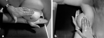
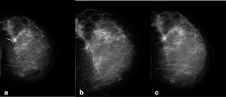
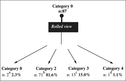
SUMMARY: BACKGROUND: The aim of this study was to investigate the efficacy of the rolled views taken in craniocaudal (CC) and mediolateral oblique (MLO) projections in solving equivocal mammography findings. PATIENTS AND METHODS: The rolled views were taken by changing the positioning of the breast but not the obliquity of the X-ray beams. The breast was rolled medially or laterally in the rolled CC view, and inferiorly or superiorly in the rolled MLO view to separate overlapping structures from each other. RESULTS: We evaluated equivocal findings in 87 asymptomatic women undergoing either CC (n = 48, 55%) or MLO (n = 39, 45%) rolled views between 2001 and 2008. The rolled views were helpful in solving equivocal mammographic findings and making proper decisions on management in 85 of the 87 (97.7%) women. This technique was used for breast asymmetries in 55 of the 87 (63.2%) women, and was sufficient to directly show summation artifacts in 59 of 79 (74.6%) women. The rolled views revealed 4 intramammary lymph nodes, 2 circumscribed masses out of 6 obscured masses, 7 summation artifacts, and 2 circumscribed masses out of 9 questionable masses. CONCLUSIONS: The rolled view is an effective method of differentiating summation artifacts from real lesions on mammography in both the CC and the MLO view.
Hintergrund: Ziel dieser Studie war die Evaluierung des Stellenwertes der gerollten Aufnahmen bei der Abklärung von unklaren mammographischen Befunden im craniocaudalen (CC) und mediolateral-schrägen (MLO) Strahlengang.
Patientinnen und Methoden: Die gerollten Aufnahmen wurden nach Veränderung der Lage der Brust und bei konstantem Röntgenstrahlengang aufgenommen. Die Brust wurde für die CC-Position nach mediolateral (gerollte CC-Aufnahme) und für die MLO-Position nach oben oder unten gerollt, um überlappende Strukturen voneinander zu trennen.
Ergebnisse: Zwischen 2001 und 2008 wurden bei 87 asymptomatischen Patientinnen 48 (55%) unklare Befunde mit einer gerollten Aufnahme in der CC-Position und 39 (45%) unklare Befunde mit einer gerollten Aufnahme in der MLO-Position evaluiert. Bei 85 der 87 Patientinnen (97,7%) führte die gerollte Aufnahme zur Abklärung des unklaren Befundes und unterstützte die Therapieentscheidung. Die Methode wurde bei 55 der 87 Patientinnen (63,2%) zur Abklärung von Brustasymmetrien eingesetzt und konnte bei 59 von 79 Patientinnen (74,6%) Summationsartefakte identifizieren. 9 suspekte Läsionen wurden mithilfe gerollter Aufnahmen evaluiert. Dabei wurden 4 intramammäre Lymphknoten, 2 umschriebene Tumoren (von 6 obskuren Neoplasien), 7 Summationsartefakte und 2 umschriebene Tumoren (von 9 fraglichen Neoplasien) aufgedeckt.
Schlussfolgerungen: Gerollte Aufnahmen sind bei der Differenzierung von Summationsartefakten und echten Läsionen sowohl in der CC-als auch der MLO-Position effektiv.
Figures
Similar articles
-
Computer-aided detection in digital mammography: comparison of craniocaudal, mediolateral oblique, and mediolateral views.Radiology. 2006 Dec;241(3):695-701. doi: 10.1148/radiol.2413051145. Radiology. 2006. PMID: 17114620
-
Correspondence in texture features between two mammographic views.Med Phys. 2005 Jun;32(6):1598-606. doi: 10.1118/1.1915013. Med Phys. 2005. PMID: 16013719
-
Are All Views with and without Displacement Maneuver Necessary in Augmentation Mammography? Putting Numbers Into Perspective.Asian Pac J Cancer Prev. 2022 Jan 1;23(1):233-239. doi: 10.31557/APJCP.2022.23.1.233. Asian Pac J Cancer Prev. 2022. PMID: 35092393 Free PMC article.
-
Breast MR Imaging for Equivocal Mammographic Findings: Help or Hindrance?Radiographics. 2016 Jul-Aug;36(4):943-56. doi: 10.1148/rg.2016150205. Epub 2016 Jun 10. Radiographics. 2016. PMID: 27284757 Review.
-
Strategies to Increase Cancer Detection: Review of True-Positive and False-Negative Results at Digital Breast Tomosynthesis Screening.Radiographics. 2016 Nov-Dec;36(7):1954-1965. doi: 10.1148/rg.2016160049. Epub 2016 Oct 7. Radiographics. 2016. PMID: 27715711 Free PMC article. Review.
References
-
- Pearson KL, Sickles EA, Frankel SD, Leung JWT. Efficacy of step-oblique mammography for confirmation and localization of densities seen on only one standard mammographie view. AJR. 2000;174:745–752. - PubMed
-
- Samardar P, Paredes ES, Grimes MM, Wilson JD. Focal asymmetric densities seen at mammography: US and pathologic correlation. Radiographies. 2002;22:19–33. - PubMed
-
- Logan WW, Janus J. Use of special mammographie views to maximize radiographie information. Radiol din North Am. 1987;25:953–959. - PubMed
-
- Sickles EA. Successful methods to reduce false-positive mammography interpretations. Radiol Clin North Am. 2000;38:693–700. - PubMed
-
- Brenner JR. False-negative mammograms: medical, legal, and risk management implications. Radiol din North Am. 2000;38:741–757. - PubMed
LinkOut - more resources
Full Text Sources
Other Literature Sources
