Case Reports
. 2010 Dec;11(4):257-61.
doi: 10.1007/s10195-010-0119-6. Epub 2010 Nov 20.

Osseous erosion by herniated nucleus pulposus mimicking intraspinal tumor: a case report

Affiliations
Case Reports

Osseous erosion by herniated nucleus pulposus mimicking intraspinal tumor: a case report

Shinji Yoshioka et al. J Orthop Traumatol. 2010 Dec.

Abstract

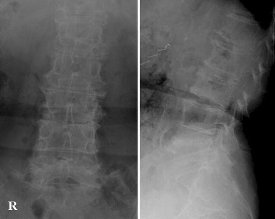
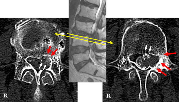
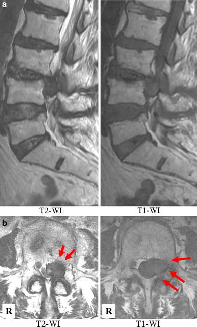
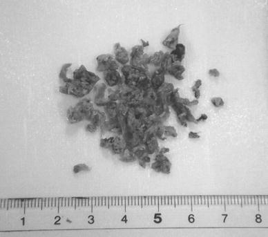
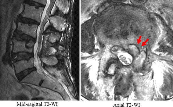
Erosion of spinal osseous structure, so-called scalloping, has been rarely reported associated with herniated nucleus pulposus (HNP). We report a rare case of HNP causing erosion of the spinal osseous structure (including lamina). The patient was an 81-year-old woman with 3-year history of low-back pain and left leg radiating pain. Muscle weakness of the left leg was also apparent. Computed tomography following myelography showed severe compression of the dural sac at the level of L3-L4; furthermore, erosion of the lamina, pedicle, and vertebral body was noted, indicating that the space-occupying mass was most probably a tumorous lesion. The mass also showed calcification inside. During the surgery, the mass was confirmed to be an HNP with calcification. Following resection, the pain disappeared. Surgeons should be aware of the possibility of scalloping of the vertebrae caused by HNP mimicking a tumorous lesion.


Figures

Fig. 1
Fig. 1
Plain AP and lateral radiographs of the lumbar spine
Fig. 2
Fig. 2
CT myelography following myelography at L3–L4 showing a tumorous mass with partial calcification in the spinal canal. The right panel shows the slice corresponding to the cranial aspect of the L4 pedicle. The left panel shows the slice corresponding to the caudal aspect of the L4 pedicle. Red arrows indicate the location of scalloping, and white arrows, the calcification in the mass
Fig. 3
Fig. 3
a MR sagittal images showing a mass at L3–L4. The mass extended caudally and strongly compressed the dural sac. b Axial MR images showing the mass located on the right side and the scalloping (red arrows)
Fig. 4
Fig. 4
The surgical specimen shown after fixation in 10% formic acid. On macroscopic examination, it seems to be an HNP
Fig. 5
Fig. 5
Postoperative MR images revealed that the thecal sac was totally decompressed. Scalloping was also clearly seen on postoperative MRI (arrows in the right panel)

Similar articles

Cited by

References

    1. El Khamary S, Alorainy IA. Case 100: spinal epidural meningioma. Radiology. 2006;241:614–617. doi: 10.1148/radiol.2412040203. - DOI - PubMed
    1. Jaiswal A, Shetty AP, Rajasekaran S. Giant cystic intradural schwannoma in the lumbosacral region: a case report. J Orthop Surg (Hong Kong) 2008;16:102–106. - PubMed
    1. Koeller KK, Rosenblum RS, Morrison AL. Neoplasms of the spinal cord and filum terminale: radiologic-pathologic correlation. Radiographics. 2000;20:1721–1749. doi: 10.1148/radiographics.20.6.g00nv151721. - DOI - PubMed
    1. Kumar R, Guinto FC, Jr, Madewell JE, Swischuk LF, David R. The vertebral body: radiographic configurations in various congenital and acquired disorders. Radiographics. 1988;8:455–485. doi: 10.1148/radiographics.8.3.3380991. - DOI - PubMed
    1. Liu JK, Cole CD, Kan P, Schmidt MH. Spinal extradural arachnoid cysts: clinical, radiological, and surgical features. Neurosurg Focus. 2007;22:E6. doi: 10.3171/foc.2007.22.2.6. - DOI - PubMed

Publication types

LinkOut - more resources