

Case Reports
doi: 10.1007/s10195-010-0116-9.
Epub 2010 Nov 20.
Pseudoaneurysm overlying an osteochondroma: a noteworthy complication
Affiliations
- PMID: 21103904
- PMCID: PMC3014466
- DOI: 10.1007/s10195-010-0116-9
Item in Clipboard
Case Reports
Pseudoaneurysm overlying an osteochondroma: a noteworthy complication
J Orthop Traumatol.
2010 Dec.
Abstract
Pseuodaneurysms are an extremely rare complication of osteochondromas. We describe a case of traumatic pseudoaneurysm of the brachial artery presenting as a soft tissue mass in a patient who was treated for an osteochondroma 3 years earlier. This case demonstrates that radiographic follow-up of large osteochondromas is mandatory and that, in patients with soft tissue masses and a history of osteochondroma, pseudoaneurysms should be included in the differential diagnosis.
Figures

Preoperative clinical image

Axial magnetic resonance image (MRI) T2 (a), T1 (b) and T1 turbo spin echo (TSE) fat-saturation (c) sequences. The mass has inhomogeneous signal on T2- (a) and T1- (b) weighted sequence, concentric aspect, regular contours (a, b) and did not diffusely enhance due to the presence of pseudoaneurysm, thrombus, and hematoma (c)
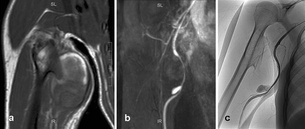
Coronal magnetic resonance imaging (MRI) T1 turbo spin echo (TSE) (a), MR angiography (b), and digital angiography (c). MR angiography (b) identifies the pseudoaneurysm arising from the compressed brachial artery, with extravasation of contrast medium into the mass, well demonstrated also by the angiography (c)
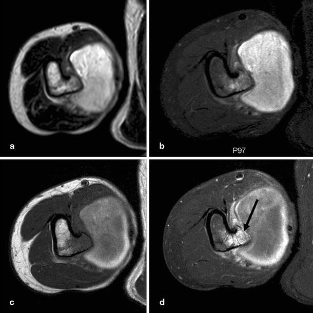
Axial magnetic resonance imaging (MRI) T2 (a), proton-density fat saturation (PD fat-sat) (b), T1 turbo spin echo (TSE) (c), T1-TSE fat-sat (d) sequences of the osteochondroma underlying the pseudoaneurysm. Note hyperintensity of the medulla adjacent to the lesion on PD sequences (b) and enhancement of the medulla itself after contrast media (arrow) (d)
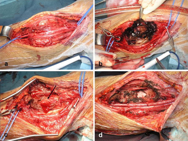
Intraoperative images (a–c). The brachial artery and the pseudoaneurysm were isolated and the artery was controlled with nylon tapes (a); the pseudoaneurysmal sac was opened and the thrombus removed (b); a 7-cm arterial segment including the pseudoaneurysm was resected, highlighting the underlying cartilaginous spur (arrow) of the osteochondroma, which probably caused the pseudoaneurysm (c); the osteochondroma was resected, and a reversed saphenous vein interposition graft was then performed (d)
References
-
- Campanacci M. Exostosis. In: Campanacci M, editor. Bone and soft tissue tumors. 2. Padova: Springer-Verlag; 1999. pp. 179–196.
-
- Stieber JR, Pierz KA, Dormans JP. Hereditary multiple exostoses: a current understanding of clinical and genetic advances. Univ PA Orthop J. 2001;14:39–48.
Publication types
MeSH terms
LinkOut - more resources
Full Text Sources
Medical
