

Catalytic residues in hydrolases: analysis of methods designed for ligand-binding site prediction
- PMID: 21104192
- PMCID: PMC3032897
- DOI: 10.1007/s10822-010-9402-0
Catalytic residues in hydrolases: analysis of methods designed for ligand-binding site prediction
Abstract
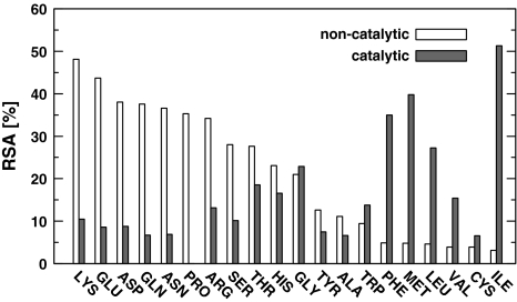
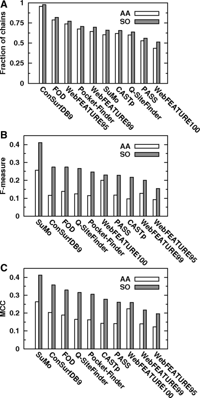
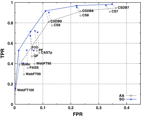
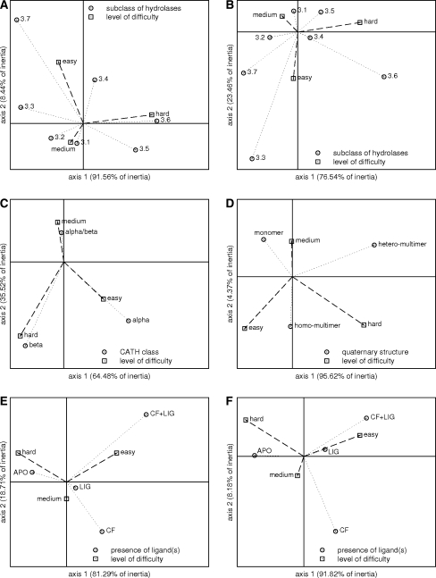
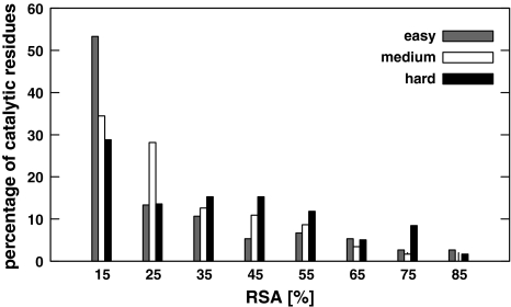
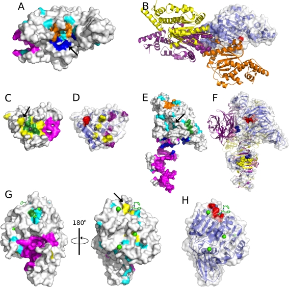
The comparison of eight tools applicable to ligand-binding site prediction is presented. The methods examined cover three types of approaches: the geometrical (CASTp, PASS, Pocket-Finder), the physicochemical (Q-SiteFinder, FOD) and the knowledge-based (ConSurf, SuMo, WebFEATURE). The accuracy of predictions was measured in reference to the catalytic residues documented in the Catalytic Site Atlas. The test was performed on a set comprising selected chains of hydrolases. The results were analysed with regard to size, polarity, secondary structure, accessible solvent area of predicted sites as well as parameters commonly used in machine learning (F-measure, MCC). The relative accuracies of predictions are presented in the ROC space, allowing determination of the optimal methods by means of the ROC convex hull. Additionally the minimum expected cost analysis was performed. Both advantages and disadvantages of the eight methods are presented. Characterization of protein chains in respect to the level of difficulty in the active site prediction is introduced. The main reasons for failures are discussed. Overall, the best performance offers SuMo followed by FOD, while Pocket-Finder is the best method among the geometrical approaches.
Figures
Similar articles
-
Q-SiteFinder: an energy-based method for the prediction of protein-ligand binding sites.Bioinformatics. 2005 May 1;21(9):1908-16. doi: 10.1093/bioinformatics/bti315. Epub 2005 Feb 8. Bioinformatics. 2005. PMID: 15701681
-
Predicting protein-ligand binding sites based on an improved geometric algorithm.Protein Pept Lett. 2011 Oct;18(10):997-1001. doi: 10.2174/092986611796378756. Protein Pept Lett. 2011. PMID: 21592081
-
MetaPocket: a meta approach to improve protein ligand binding site prediction.OMICS. 2009 Aug;13(4):325-30. doi: 10.1089/omi.2009.0045. OMICS. 2009. PMID: 19645590
-
Bioinformatics and molecular modeling in chemical enzymology. Active sites of hydrolases.Biochemistry (Mosc). 2002 Oct;67(10):1099-108. doi: 10.1023/a:1020907122341. Biochemistry (Mosc). 2002. PMID: 12460108 Review.
-
Protein-protein interface analysis and hot spots identification for chemical ligand design.Curr Pharm Des. 2014;20(8):1192-200. doi: 10.2174/13816128113199990065. Curr Pharm Des. 2014. PMID: 23713772 Review.
Cited by
-
Structural Specificity of Polymorphic Forms of α-Synuclein Amyloid.Biomedicines. 2023 Apr 29;11(5):1324. doi: 10.3390/biomedicines11051324. Biomedicines. 2023. PMID: 37238996 Free PMC article.
-
A new protein-ligand binding sites prediction method based on the integration of protein sequence conservation information.BMC Bioinformatics. 2011 Dec 14;12 Suppl 14(Suppl 14):S9. doi: 10.1186/1471-2105-12-S14-S9. BMC Bioinformatics. 2011. PMID: 22373099 Free PMC article.
-
Internal force field in proteins seen by divergence entropy.Bioinformation. 2011;6(8):300-2. doi: 10.6026/97320630006300. Epub 2011 Jul 6. Bioinformation. 2011. PMID: 21769190 Free PMC article.
-
External Force Field for Protein Folding in Chaperonins-Potential Application in In Silico Protein Folding.ACS Omega. 2024 Apr 10;9(16):18412-18428. doi: 10.1021/acsomega.4c00409. eCollection 2024 Apr 23. ACS Omega. 2024. PMID: 38680295 Free PMC article.
-
Intermediates in the protein folding process: a computational model.Int J Mol Sci. 2011;12(8):4850-60. doi: 10.3390/ijms11084850. Epub 2011 Jul 29. Int J Mol Sci. 2011. PMID: 21954329 Free PMC article.
References
-
- Brenner SE. A tour of structural genomics. Nat Rev Genet. 2001;2:801–809. - PubMed
-
- Chandonia J-M, Brenner SE. The impact of structural genomics: expectations and outcomes. Science. 2006;311:347–351. - PubMed
-
- Gileadi O, Knapp S, Lee WH, Marsden BD, Müller S, Niesen FH, Kavanagh KL, Ball LJ, von Delft F, Doyle DA, Oppermann UCT, Sundström M. The scientific impact of the structural genomics consortium: a protein family and ligand-centered approach to medically-relevant human proteins. J Struct Funct Genomics. 2007;8:107–119. - PMC - PubMed
-
- Hajduk PJ, Huth JR, Tse C. Predicting protein druggability. Drug Discov Today. 2005;10:1675–1682. - PubMed
Publication types
MeSH terms
Substances
LinkOut - more resources
Full Text Sources
