Review
. 2011 Mar;68(6):1011-20.
doi: 10.1007/s00018-010-0595-0. Epub 2010 Nov 23.

Modulation of E-cadherin function and dysfunction by N-glycosylation

Affiliations
Review

Modulation of E-cadherin function and dysfunction by N-glycosylation

Salomé S Pinho et al. Cell Mol Life Sci. 2011 Mar.

Abstract

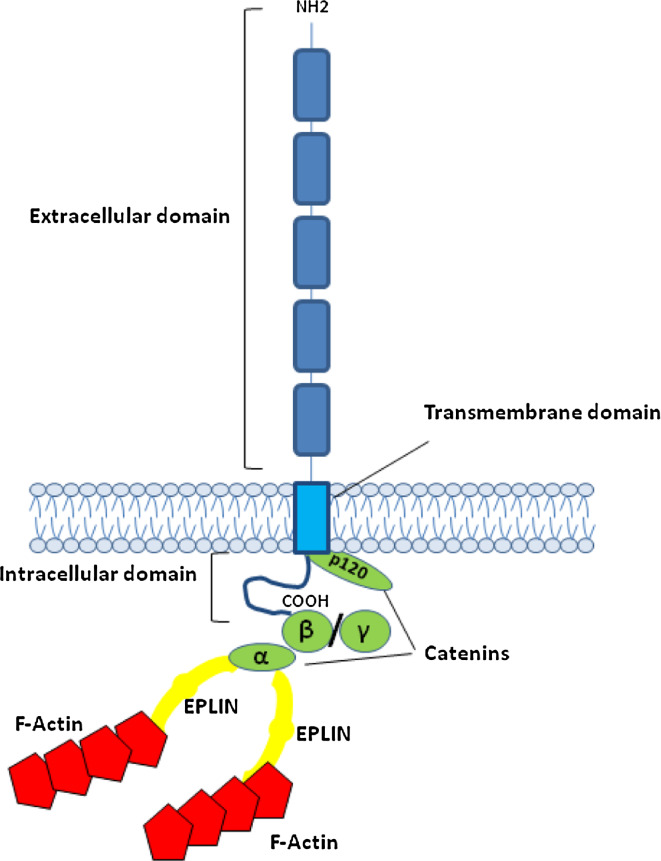
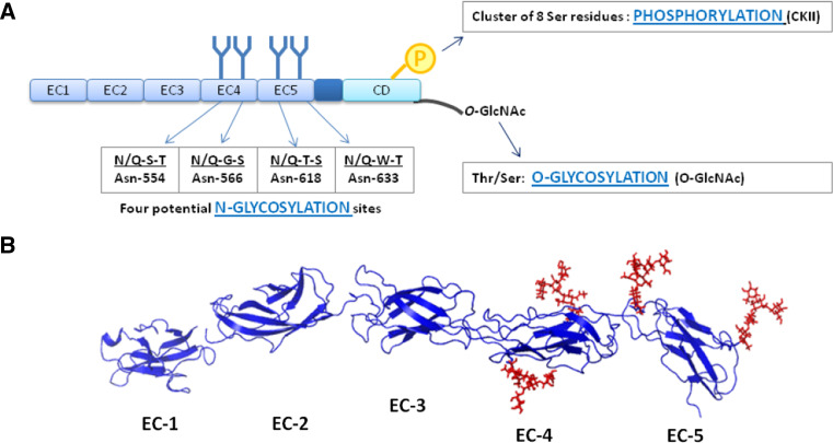
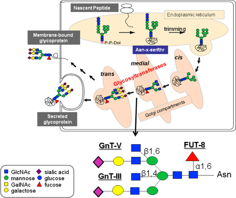
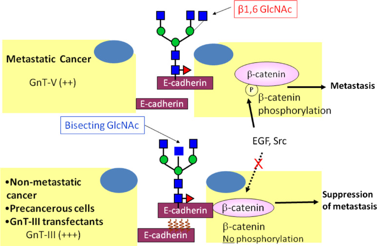
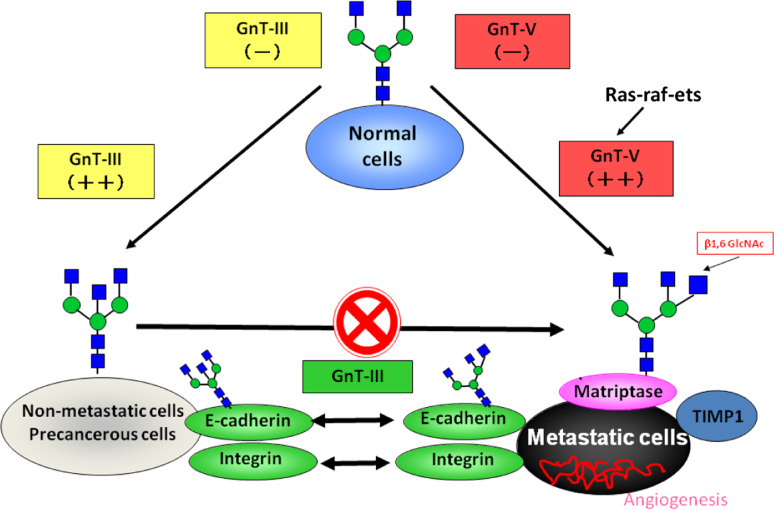
Several mechanisms have been proposed to explain the E-cadherin dysfunction in cancer, including genetic and epigenetic alterations. Nevertheless, a significant number of human carcinomas have been seen that show E-cadherin dysfunction that cannot be explained at the genetic/epigenetic level. A substantial body of evidence has appeared recently that supports the view that other mechanisms operating at the post-translational level may also affect E-cadherin function. The present review addresses molecular aspects related to E-cadherin N-glycosylation and evidence is presented showing that the modification of N-linked glycans on E-cadherin can affect the adhesive function of this adhesion molecule. The role of glycosyltransferases involved in the remodeling of N-glycans on E-cadherin, including N-acetylglucosaminyltransferase III (GnT-III), N-acetylglucosaminyltransferase V (GnT-V), and the α1,6 fucosyltransferase (FUT8) enzyme, is also discussed. Finally, this review discusses an alternative functional regulatory mechanism for E-cadherin operating at the post-translational level, N-glycosylation, that may underlie the E-cadherin dysfunction in some carcinomas.


Figures

Fig. 1
Fig. 1
Schematic representation of the E-cadherin–catenin complex. The E-cadherin–catenin complex is proposed to interact with F-actin via α-catenin association with actin-binding proteins such as EPLIN [5]. β-Catenin and γ-catenin bind to E-cadherin in a mutually exclusive manner
Fig. 2
Fig. 2
E-cadherin post-translational modifications. a The extracellular domain (EC) of human E-cadherin contains four potential N-glycosylation sites, which are located in EC4 and EC5. The phosphorylation of E-cadherin by casein kinase II (CKII) can occur in a short stretch of 30 aa in the cytoplasmic domain (CD), which contains a cluster of 8 Ser residues. Cytoplasmic O-glycosylation (O-GlcNAc addition in Thr/Ser residues) has been reported to regulate E-cadherin. These mechanims can modulate E-cadherin mediated cell–cell adhesion at a post-translational level. b Three-dimensional structure of the extracellular domain (EC1–EC5) of E-cadherin. The crystal structure of human EC1 was used in this representation and EC2–EC5 were modeled based on the crystal structure of C-cadherin. Four N-glycans were modeled with GlyProt (http://www.glycosciences.de/glyprot/), as shown in red
Fig. 3
Fig. 3
Biosynthesis of N-linked glycans. Representation of a N-glycan structure with the reactions catalyzed by GnT-III, GnT-V, and FUT8
Fig. 4
Fig. 4
Regulatory mechanism of E-cadherin-mediated cell–cell adhesion and GnT-III/GnT-V. GnT-III activity is associated with an increase in bisecting GlcNAc structures in E-cadherin, leading to a concomitant decrease in β1,6 branched structures, due to competition with GnT-V glycosyltransferase. The addition of bisecting GlcNAc residues to E-cadherin down-regulates the tyrosine phosphorylation of β-catenin and thus enhances cell–cell binding to suppress metastasis (lower figure). Conversely, in metastatic cancer cells (upper figure), the addition of β1,6 branched structures by GnT-V to E-cadherin is associated with increased tyrosine phosphorylation of β-catenin through the EGFR and Src signaling pathways, and therefore reduces E-cadherin-mediated cell–cell adhesion thereby contributing to the promotion of cancer metastasis
Fig. 5
Fig. 5
The role of N-glycan structures in the carcinogenic process. In normal cells, the GnT-III and GnT-V enzymes are normally underexpressed. The overexpression of GnT-III is associated with increased synthesis of bisecting GlcNAc structures in some important target glycoproteins involved in cell adhesion such as E-cadherin and integrins, the modification of which by bisecting N-glycans is associated with the suppression of metastasis through enhancement of E-cadherin-mediated cell–cell adhesion and a decrease in integrin-mediated cell-extracellular matrix adhesion. Furthermore, GnT-III up-regulation precludes the availability of the substrate for the GnT-V enzyme, which is no longer able to synthesize branched structures. In a metastatic cancer situation, activation of the ras-raf-ets signaling pathway regulates the transcription of the GnT-V gene and the resulting increase in GnT-V leads to increased enzymatic production of β1,6 branched structures that modify glycoproteins involved in the carcinogenic process, including Matriptase; TIMP-1 (Tissue Inhibitor of Metalloproteinase-1, in which β1,6 branching correlates with the invasive and metastatic potential of cancer cells) as well as integrins and E-cadherin, the modification of which contributes to a decrease in cell–cell adhesion, and increase in tumor cell invasion and migration. In addition, other mechanisms also indicate that a secreted type of GnT-V may contribute to tumor angiogenesis

Similar articles

Cited by

References

    1. Gumbiner BM. Regulation of cadherin-mediated adhesion in morphogenesis. Nat Rev Mol Cell Biol. 2005;6:622–634. doi: 10.1038/nrm1699. - DOI - PubMed
    1. Tamura K, Shan WS, Hendrickson WA, Colman DR, Shapiro L. Structure–function analysis of cell adhesion by neural (N-) cadherin. Neuron. 1998;20:1153–1163. doi: 10.1016/S0896-6273(00)80496-1. - DOI - PubMed
    1. Shi QM, Maruthamuthu V, Li F, Leckband D. Allosteric cross talk between cadherin extracellular domains. Biophys J. 2010;99:95–104. doi: 10.1016/j.bpj.2010.03.062. - DOI - PMC - PubMed
    1. Nelson WJ. Regulation of cell–cell adhesion by the cadherin–catenin complex. Biochem Soc Trans. 2008;36:149–155. doi: 10.1042/BST0360149. - DOI - PMC - PubMed
    1. Abe K, Takeichi M. EPLIN mediates linkage of the cadherin catenin complex to F-actin and stabilizes the circumferential actin belt. Proc Natl Acad Sci U S A. 2008;105:13–19. doi: 10.1073/pnas.0710504105. - DOI - PMC - PubMed

Publication types

LinkOut - more resources