Review
. 2011 Feb;28(2):237-59.
doi: 10.1007/s11095-010-0318-0. Epub 2010 Nov 23.

Inorganic nanoparticles in cancer therapy

Affiliations
Review

Inorganic nanoparticles in cancer therapy

Sanjib Bhattacharyya et al. Pharm Res. 2011 Feb.

Abstract

Nanotechnology is an evolving field with enormous potential for biomedical applications. The growing interest to use inorganic nanoparticles in medicine is due to the unique size- and shape-dependent optoelectronic properties. Herein, we will focus on gold, silver and platinum nanoparticles, discussing recent developments for therapeutic applications with regard to cancer in terms of nanoparticles being used as a delivery vehicle as well as therapeutic agents. We will also discuss some of the key challenges to be addressed in future studies.


Figures

Figure 1
Figure 1
Left: Transmission electron micrographs of Au nanospheres and nanorods (a, b) and Ag nanoprisms (c, mostly truncated triangles) formed using citrate reduction, seeded growth, and DMF reduction, respectively. Right: Photographs of colloidal dispersions of Au-Ag alloy nanoparticles with increasing Au concentration (d), Au nanorods of increasing aspect ratio (e), and Ag nanoprisms with increasing lateral size (f). Reprinted with permission from Ref [40], Marzan et al., Nanometals: Formation and color. Materials Today. 7, 26 (2004). Copyright © 2004, Elsevier Limited.
Figure 2
Figure 2
In vivo fluorescence images of tumor-bearing mice using Qdot probes with anti-PEG-PSMA antibody conjugates surface modifications in mouse cancer model (PSMA: Prostate specific membrane antigen). In vivo fluorescence imaging was carried out using a macroillumination system. Reprinted with permission from Ref [57], Gao et al., In vivo cancer targeting and imaging with semiconductor quantum dots. Nat Biotech. 22, 969 (2004). Copyright © 2004, Nature publishing group.
Figure 3
Figure 3
Laser scanning confocal reflectance images of (a) precancerous and (b) normal fresh cervical ex vivo tissue labeled with anti-EGFR/Au nanoparticle bioconjugates. The images were obtained with 647 nm excitation wavelength, and are false-colored in red. Reprinted with permission from Ref [62], Sokolov et al., Real-Time Vital Optical Imaging of Precancer Using Anti-Epidermal Growth Factor Receptor Antibodies Conjugated to Gold Nanoparticles. Cancer Res. 63, 1999 (2003). Copyright © 2003, American Association for Cancer Research.
Figure 4
Figure 4
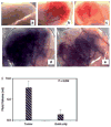
Polymeric nanoparticles are shown as representative nanocarriers (circles). Passive tissue targeting is achieved by extravasation of nanoparticles through increased permeability of the tumour vasculature and ineffective lymphatic drainage (EPR effect). Active cellular targeting (inset) can be achieved by functionalizing the surface of nanoparticles with ligands that promote cell-specific recognition and binding. The nanoparticles can (i) release their contents in close proximity to the target cells; (ii) attach to the membrane of the cell and act as an extracellular sustained-release drug depot; or (iii) internalize into the cell. Reprinted with permission from Ref [73], Langer et al. Nanocarriers as an emerging platform for cancer therapy. Nature Nanotechnology 2, 751 (2007). © 2010 Nature Publishing Group. Figure 4A: In vivo targeting of the nanoconjugate and its therapeutic efficacy. A, the quantification of the amount of gold taken up by the tumor, kidney, and liver under different treatment groups (n = 3). A comparative bioluminescence image from the mice treated with a mixture of C225 and gemcitabine (C225 + Gem; B) or Au-C225-Gem (C) i.p. (n = 10). D, effect of different treatment groups on tumor growth inhibition in vivo (left). Tumor volume was measured after sacrificing the mice at the end of the experiment. Right, plasma concentration of gold over time determined by ICP analysis. Blood samples were collected from the mice under isoflurane anesthesia at different time points in heparinized tubes containing tetrahydrouridine to prevent gemcitabine degradation by cytidine deaminase after i.v. drug administration. Reprinted with permission from Ref [76], Patra et al., Selective laser photo-thermal therapy of epithelial carcinoma using anti-EGFR antibody conjugated gold nanoparticles. Cancer Res. 68, 1970 (2008). Copyright © 2008, American Association for Cancer Research.
Figure 4
Figure 4
Polymeric nanoparticles are shown as representative nanocarriers (circles). Passive tissue targeting is achieved by extravasation of nanoparticles through increased permeability of the tumour vasculature and ineffective lymphatic drainage (EPR effect). Active cellular targeting (inset) can be achieved by functionalizing the surface of nanoparticles with ligands that promote cell-specific recognition and binding. The nanoparticles can (i) release their contents in close proximity to the target cells; (ii) attach to the membrane of the cell and act as an extracellular sustained-release drug depot; or (iii) internalize into the cell. Reprinted with permission from Ref [73], Langer et al. Nanocarriers as an emerging platform for cancer therapy. Nature Nanotechnology 2, 751 (2007). © 2010 Nature Publishing Group. Figure 4A: In vivo targeting of the nanoconjugate and its therapeutic efficacy. A, the quantification of the amount of gold taken up by the tumor, kidney, and liver under different treatment groups (n = 3). A comparative bioluminescence image from the mice treated with a mixture of C225 and gemcitabine (C225 + Gem; B) or Au-C225-Gem (C) i.p. (n = 10). D, effect of different treatment groups on tumor growth inhibition in vivo (left). Tumor volume was measured after sacrificing the mice at the end of the experiment. Right, plasma concentration of gold over time determined by ICP analysis. Blood samples were collected from the mice under isoflurane anesthesia at different time points in heparinized tubes containing tetrahydrouridine to prevent gemcitabine degradation by cytidine deaminase after i.v. drug administration. Reprinted with permission from Ref [76], Patra et al., Selective laser photo-thermal therapy of epithelial carcinoma using anti-EGFR antibody conjugated gold nanoparticles. Cancer Res. 68, 1970 (2008). Copyright © 2008, American Association for Cancer Research.
Figure 5
Figure 5
Schematic drawing illustrating the concept of folate targeting of liposomes to tumor cells. The blue dots represent the liposomal folate ligands. The red dots represent the drug molecules encapsulated in the liposome water phase. The various steps involved in the targeting process are numerically designated from 1 to 6. Steps 1–3 are common to nontargeted and targeted liposomes. Steps 4–6 are specific to FTL. (1) Liposomes with long-circulating properties increase the number of passages through the tumor microvasculature. (2) Increased vascular permeability in tumor tissue enables properly downsized liposomes to extravasate and reach the tumor interstitial fluid. (3) Drug is gradually released from liposomes remaining in the interstitial fluid and enters tumor cells as free drug to exert a cytotoxic effect. (4) Other liposomes bind to the FR expressed on the tumor cell membrane via the folate ligand. Because of the limited diffusion capacity of liposomes, binding is likely to be limited to those tumor cells in closest vicinity to blood vessels. (5) Liposomes are internalized by tumor cells via FRME. (6) Internalized liposomes release their drug content in the cytosol enabling the drug to exert its cytotoxic effect. Reprinted with permission from Ref [81], Ratnam et al. Tumor cell targeting of liposome-entrapped drugs with phospholipid-anchored folic acid–PEG conjugates. Adv Drug Deliv Rev. 56, 1177 (2004). ©2004, Elsevier Limited.
Figure 6
Figure 6
Effect of nanogold on angiogenesis in vivo in the ears of nude mice. Gross appearance of angiogenesis 7 days (see Materials and Methods) after injection of nanogold only (A), Ad-VEGF only (B), nanogold and Ad-VEGF (C). Giemsa stained 1 μmol/L epon sections of the ears were photographed at 10× magnifications, nanogold only. (G), nanogold and Ad-VEGF (H), ascites fluid accumulation in the peritoneal cavity (I). Reprinted with permission from [14], Mukherjee et al. Antiangiogenic Properties of Gold Nanoparticles. Clin Cancer Res. 11, 3530 (2005). ©2004, American Association for Cancer Research.
Figure 7
Figure 7
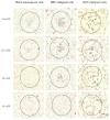
Light scattering images of HaCaT benign cells (left), HSC malignant cells (middle) and HOC malignant cells (right) after incubation with anti-EGFR antibody conjugated gold nanoparticles. The figure shows clearly distinguished difference for the scattering images between the benign and malignant cells. The conjugated nanoparticles bind specifically with high concentrations to the surface of the malignant cells. Scale bar: 10 μm for all images. HaCaT benign cells (top row), HSC malignant cells (middle row) and HOC malignant cells (bottom row) irradiated at different laser powers and then stained with trypan blue. HaCaT benign cells were killed at and above 57 W/cm2, HSC malignant cells were killed at and above 25 W/cm2 and HOC malignant cells were killed at and above 19 W/cm2. Scale bar: 60 μm for all images (For interpretation of the reference to color in this legend, the reader is referred to the web version of this article). Reprinted with permission from Ref [118], I.H. El-Sayed et al., Selective laser photo-thermal therapy of epithelial carcinoma using anti-EGFR antibody conjugated gold nanoparticles. Cancer Lett. 239, 129 (2006). Copyright © 2006, Elsevier limited.
Figure 8
Figure 8
Selective photothermal therapy of cancer cells with anti-EGFR/Au nanorods incubated. The circles show the laser spots on the samples. At 80 mW (10 W/cm2), the HSC and HOC malignant cells are obviously injured while the HaCat normal cells are not affected. The HaCat normal cells start to be injured at 120 mW (15 W/cm2) and are obviously injured at 160 mW (20 W/cm2). Reprinted with permission from Ref [70], H. Huang et al., Cancer Cell Imaging and Photothermal Therapy in the Near-Infrared Region by Using Gold Nanorods. J. Am. Chem. Soc. 128, 2115 (2006). © 2006, American Chemical Society.
Figure 9
Figure 9
Fast processes (t < 10–12 s) involved in platinum nanoparticles excited by ionizing radiations. Reprinted with permission from Ref [163], Porcel et al. Platinum nanoparticles: a promising material for future cancer therapy. Nanotechnology. 21, 1 (2010). Copyright © 2010, IOP publishing limited.
Fig. 10
Fig. 10
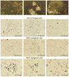
Apoptosis of A549 cells incubated with nanoparticles: LMH (A), iron oxide (B), silica (C), SWCNT (D), and % apoptotic cells (E). Cells (2 × 10⁴ cells/ml) were exposed to nanoparticles (250–500 lg/ml) for 72 h and apoptotic cells were measured by annexin V-FITC (green) binding assay. The nuclei were stained with Dapi (blue) or PI (red). Cell membrane partially stained by annexin V-FITC was indicated by the arrow. *Significant difference from control (p < 0.05). Reprinted with permission from Ref [196], Choi et al., Toxicological effects of inorganic nanoparticles on human lung cancer A549 cells. J. Inorg. Biochem. 239, 129 (2009). Copyright © 2006, Elsevier limited.
Fig. 11
Fig. 11
A proposed mechanism of MNP-induced macrophage recruitment into neuronal tissues. (1) Exposure to cytotoxic MNPs stimulated the formation of ROS in resident cells.(2) ROS promotes expression and release of proinflammatory cytokines, such as TNF-α. Through its two receptors (TNFR), TNF-α activates p38 and ERK mitogen-activated protein kinases pathways to (3) induce the expression of matrix metalloproteinases (MMPs) in its inactive, pro-MMP form. In addition, (4) ROS can directly promote MMP activation from pro-form. MMPs are the only enzymes in the body capable of degrading blood–brain and blood–nerve barriers (BBB/BNB), which (5) promotes infiltration of circulatingmacrophages (mΦ) into neuronal tissues. MNP size and surface chemistry determines the mechanisms and the target cells of MNP internalization, as well as extent of neurotoxicity of MNPs. Reprinted with permission from Ref [54], Shubayev et al., Magnetic nanoparticles for theragnostics. Adv Drug Del Rev. 61, 467 (2009). Copyright © 2009, Elsevier limited.

Similar articles

Cited by

References

    1. Giljohannand DA, Mirkin CA. Drivers of biodiagnostic development. Nature. 2009;462:461–464. - PMC - PubMed
    1. Patra CR, Bhattacharya R, Mukhopadhyay D, Mukherjee P. Application of gold nanoparticles for targeted therapy in cancer. J Biomed Nanotechnol. 2008;4:99–132.
    1. Paciotti GF, Myer L, Weinreich D, Goia D, Pavel N, McLaughlin RE, Tamarkin L. Colloidal gold: A novel nanoparticle vector for tumor directed drug delivery. Drug Deliv. 2004;11:169–183. - PubMed
    1. Mahdihassan S. Cinnabar-gold as the best alchemical drug of longevity, called Makaradhwaja in India. Am J Chin Med. 1985;13:93–108. - PubMed
    1. Danieland D, Astruc MC. Gold Nanoparticles: Assembly, Supramolecular Chemistry, Quantum-Size-Related Properties, and Applications Toward Biology, Catalysis, and Nanotechnology. Chem Rev. 2004;104:293–346. - PubMed

Publication types