

Neuronal calcium sensor-1 regulation of calcium channels, secretion, and neuronal outgrowth
- PMID: 21104311
- PMCID: PMC11498851
- DOI: 10.1007/s10571-010-9588-7
Neuronal calcium sensor-1 regulation of calcium channels, secretion, and neuronal outgrowth
Abstract
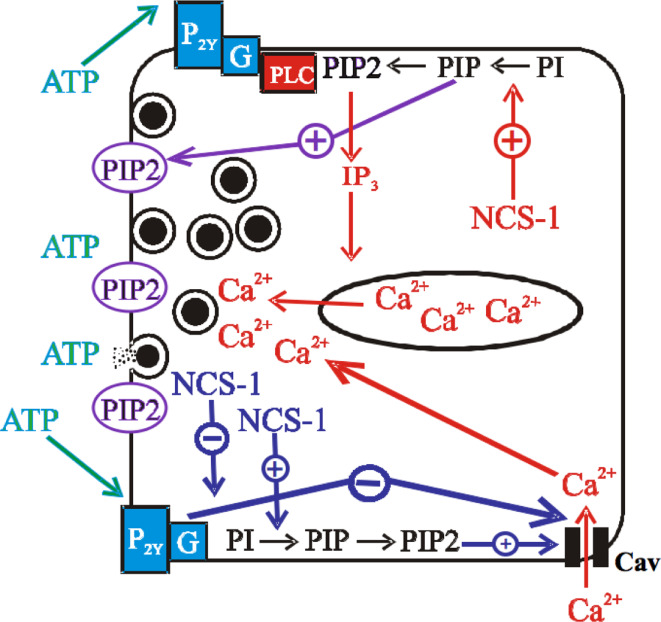
Calcium (Ca(2+)) is an important intracellular messenger underlying cell physiology. Ca(2+) channels are the main entry route for Ca(2+) into excitable cells, and regulate processes such as neurotransmitter release and neuronal outgrowth. Neuronal Calcium Sensor-1 (NCS-1) is a member of the Calmodulin superfamily of EF-hand Ca(2+) sensing proteins residing in the subfamily of NCS proteins. NCS-1 was originally discovered in Drosophila as an overexpression mutant (Frequenin), having an increased frequency of Ca(2+)-evoked neurotransmission. NCS-1 is N-terminally myristoylated, can bind intracellular membranes, and has a Ca(2+) affinity of 0.3 μM. Over 10 years ago it was discovered that NCS-1 overexpression enhances Ca(2+)-evoked secretion in bovine adrenal chromaffin cells. The mechanism was unclear, but there was no apparent direct effect on the exocytotic machinery. It was revealed, again in chromaffin cells, that NCS-1 regulates voltage-gated Ca(2+) channels (Cavs) in G-Protein Coupled Receptor (GPCR) signaling pathways. This work in chromaffin cells highlighted NCS-1 as an important modulator of neurotransmission. NCS-1 has since been shown to regulate and/or directly interact with many proteins including Cavs (P/Q, N, and L), TRPC1/5 channels, GPCRs, IP3R, and PI4 kinase type IIIβ. NCS-1 also affects neuronal outgrowth having roles in learning and memory affecting both short- and long-term synaptic plasticity. It is not known if NCS-1 affects neurotransmission and synaptic plasticity via its effect on PIP2 levels, and/or via a direct interaction with Ca(2+) channels or their signaling complexes. This review gives a historical account of NCS-1 function, examining contributions from chromaffin cells, PC12 cells and other models, to describe how NCS-1's regulation of Ca(2+) channels allows it to exert its physiological effects.
Figures
Similar articles
-
Neuronal Ca(2+) sensor 1. Characterization of the myristoylated protein, its cellular effects in permeabilized adrenal chromaffin cells, Ca(2+)-independent membrane association, and interaction with binding proteins, suggesting a role in rapid Ca(2+) signal transduction.J Biol Chem. 1999 Oct 15;274(42):30258-65. doi: 10.1074/jbc.274.42.30258. J Biol Chem. 1999. PMID: 10514519
-
Alterations in exocytosis induced by neuronal Ca2+ sensor-1 in bovine chromaffin cells.J Neurosci. 2002 Apr 1;22(7):2427-33. doi: 10.1523/JNEUROSCI.22-07-02427.2002. J Neurosci. 2002. PMID: 11923406 Free PMC article.
-
Demonstration of binding of neuronal calcium sensor-1 to the cav2.1 p/q-type calcium channel.Biochemistry. 2014 Sep 30;53(38):6052-62. doi: 10.1021/bi500568v. Epub 2014 Sep 15. Biochemistry. 2014. PMID: 25188201 Free PMC article.
-
Neuronal calcium sensor-1: a multifunctional regulator of secretion.Biochem Soc Trans. 2003 Aug;31(Pt 4):828-32. doi: 10.1042/bst0310828. Biochem Soc Trans. 2003. PMID: 12887315 Review.
-
Emerging Roles of Neuronal Ca2+ Sensor-1 in Cardiac and Neuronal Tissues: A Mini Review.Front Mol Neurosci. 2019 Mar 4;12:56. doi: 10.3389/fnmol.2019.00056. eCollection 2019. Front Mol Neurosci. 2019. PMID: 30886571 Free PMC article. Review.
Cited by
-
Control of Neuronal Ryanodine Receptor-Mediated Calcium Signaling by Calsenilin.Mol Neurobiol. 2019 Jan;56(1):525-534. doi: 10.1007/s12035-018-1080-2. Epub 2018 May 5. Mol Neurobiol. 2019. PMID: 29730765 Free PMC article.
-
Interaction of ARF-1.1 and neuronal calcium sensor-1 in the control of the temperature-dependency of locomotion in Caenorhabditis elegans.Sci Rep. 2016 Jul 20;6:30023. doi: 10.1038/srep30023. Sci Rep. 2016. PMID: 27435667 Free PMC article.
-
Dimerization of Neuronal Calcium Sensor Proteins.Front Mol Neurosci. 2018 Nov 2;11:397. doi: 10.3389/fnmol.2018.00397. eCollection 2018. Front Mol Neurosci. 2018. PMID: 30450035 Free PMC article. Review.
-
Long-term continuous corticosterone treatment decreases VEGF receptor-2 expression in frontal cortex.PLoS One. 2011;6(5):e20198. doi: 10.1371/journal.pone.0020198. Epub 2011 May 27. PLoS One. 2011. PMID: 21647420 Free PMC article.
-
NCS-1 protein regulates TRPA1 channel through the PI3K pathway in breast cancer and neuronal cells.J Physiol Biochem. 2024 May;80(2):451-463. doi: 10.1007/s13105-024-01016-z. Epub 2024 Apr 2. J Physiol Biochem. 2024. PMID: 38564162 Free PMC article.
References
-
- Aldea M, Jun K, Shin HS, Andres-Mateos E, Solis-Garrido LM, Montiel C, Garcia AG, Albillos A (2002) A perforated patch-clamp study of calcium currents and exocytosis in chromaffin cells of wild-type and α1A knockout mice. J Neurochem 81:911–921 - PubMed
-
- Angaut-Petit D, Toth P, Rogero O, Faille L, Tejedor FJ, Ferrus A (1998) Enhanced neurotransmitter release is associated with reduction of neuronal branching in a Drosophila mutant overexpressing Frequenin. Eur J Neurosci 10:423–434 - PubMed
-
- Beech DJ, Bernheim L (1992) Pertussis toxin and voltage dependence distinguish multiple pathways modulating calcium channels of rat sympathetic neurons. Neuron 8:97–106 - PubMed
-
- Bezzerides VJ, Ramsey IS, Kotecha S, Greka A, Clapham DE (2004) Rapid vesicular translocation and insertion of TRP channels. Nat Cell Biol 6(8):709–720 - PubMed
Publication types
MeSH terms
Substances
LinkOut - more resources
Full Text Sources
Molecular Biology Databases
Miscellaneous
