. 2010 Dec;42(6):871-80.
doi: 10.1002/mus.21764.

Progressive resistance voluntary wheel running in the mdx mouse

Affiliations

Progressive resistance voluntary wheel running in the mdx mouse

Jarrod A Call et al. Muscle Nerve. 2010 Dec.

Abstract

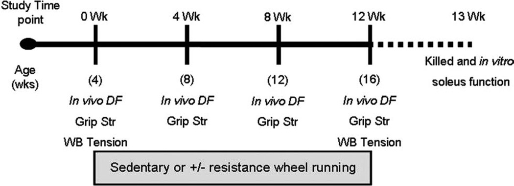
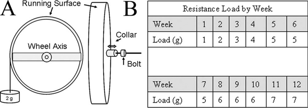
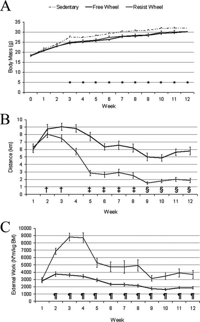
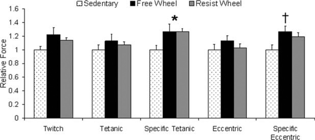
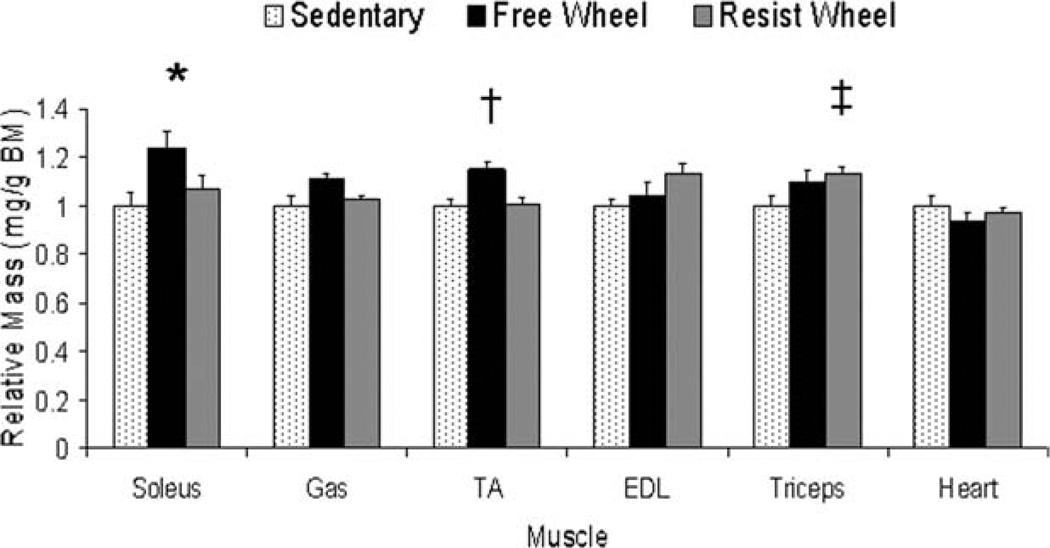
Exercise training has been minimally explored as a therapy to mitigate the loss of muscle strength for individuals with Duchenne muscular dystrophy (DMD). Voluntary wheel running is known to elicit beneficial adaptations in the mdx mouse model for DMD. The aim of this study was to examine progressive resistance wheel running in mdx mice by comprehensively testing muscle function before, during, and after a 12-week training period. Male mdx mice at ~4 weeks age were randomized into three groups: Sedentary, Free Wheel, and Resist Wheel. Muscle strength was assessed via in vivo dorsiflexion torque, grip strength, and whole body tension intermittently throughout the training period. Contractility of isolated soleus muscles was analyzed at the study's conclusion. Both Free and Resist Wheel mice had greater grip strength (~22%) and soleus muscle specific tetanic force (26%) compared with Sedentary mice. This study demonstrates that two modalities of voluntary exercise are beneficial to dystrophic muscle and may help establish parameters for an exercise prescription for DMD.


Figures

FIGURE 1
FIGURE 1
After an initial in vivo strength assessment of dorsiflexion torque (in vivo DF), grip strength (Grip Str), and whole body tension (WB Tension), mice were randomly assigned to either the Sedentary, Free Wheel, or Resist Wheel group (0 Wk). The mice assigned to the Free and Resist Wheel groups initiated running at this time-point. At the 4- and 8-week time-points, all mice were reassessed for in vivo dorsiflexion torque and grip strength. At week 12, all mice were assessed for in vivo dorsiflexion, grip strength, and whole body tension. All mice were killed 1 week later, and skeletal muscles were harvested for in vitro contractility and injury assessment, muscle mass, and Western blot analysis.
FIGURE 2
FIGURE 2
Resistance wheel design and resistance load during the 12-week training period. (A) To calibrate the resistance on the wheel a mass (e.g., 2 g) was placed on the horizontal moment arm of the wheel. The metal collar was then tightened (↓) against the axis of the wheel to maintain the resistance during wheel movement. When the appropriate resistance was reached, a bolt was tightened against the collar to secure its position. The calibration mass was then removed from the horizontal moment arm. (B) Resistance load applied to the wheel of Resist Wheel mice increased progressively from week 1 to week 12.
FIGURE 3
FIGURE 3
Body mass, averaged daily running distance, and averaged daily external work varied among groups and weeks during the 12-week study. (A) Starting at week 3, body mass was significantly greater in Sedentary mice compared with Free and Resist Wheel mice and stayed elevated for the remainder of the study (*Sedentary > Free and Resist Wheel). (B) Independent of group, averaged daily distance peaked at week 2 (†>week-1), then decreased at week 5 (‡<weeks 2 and 3) and week 9 (§ <weeks 1, 2, 3, and 4). (C) Averaged daily external work was greater at week 2 in Resist Wheel mice and stayed greater throughout the remainder of the study (¶>Free Wheel).
FIGURE 4
FIGURE 4
Free and resistance voluntary wheel running improved soleus muscle force. Data are shown relative to Sedentary values (*Free Wheel and Resist Wheel > Sedentary; †Free Wheel > Sedentary).
FIGURE 5
FIGURE 5
Hindlimb [soleus, gastrocnemius (Gas), tibialis anterior (TA), extensor digitorum longus (EDL), and forelimb (triceps)] muscle masses were affected differently by free and resistance voluntary wheel running. Data are shown relative to Sedentary values (*Free Wheel > Sedentary; †Free Wheel > Sedentary and Resist Wheel; ‡Resist Wheel > Sedentary).
FIGURE 6
FIGURE 6
Cytoskeletal protein expression in soleus muscle. (A) Representative blots of talin (269 kDa), α7-integrin (125 kDa), vinculin (117 kDa), and β-dystroglycan (B-DG, 43 kDa). Lane S is Sedentary, lane F is Free Wheel, and lane R is Resist Wheel. In addition, a representative Ponceau stain is shown verifying that equal amounts of total protein were loaded into each well. (B) Vinculin content was greater for Free Wheel mice compared with Sedentary mice in the soleus muscle (*). Values are represented relative to Sedentary.

References

    1. Aitkens SG, McCrory MA, Kilmer DD, Bernauer EM. Moderate resistance exercise program: its effect in slowly progressive neuromuscular disease. Arch Phys Med Rehabil. 1993;74:711–715. - PubMed
    1. Lynch GS, Hinkle RT, Faulkner JA. Force and power output of diaphragm muscle strips from mdx and control mice after clenbuterol treatment. Neuromuscul Disord. 2001;11:192–196. - PubMed
    1. Sacco P, Jones DA, Dick JR, Vrbova G. Contractile properties and susceptibility to exercise-induced damage of normal and mdx mouse tibialis anterior muscle. Clin Sci (Lond) 1992;82:227–236. - PubMed
    1. Petrof BJ, Shrager JB, Stedman HH, Kelly AM, Sweeney HL. Dystrophin protects the sarcolemma from stresses developed during muscle contraction. Proc Natl Acad Sci USA. 1993;90:3710–3714. - PMC - PubMed
    1. Grange RW, Call JA. Recommendations to define exercise prescription for Duchenne muscular dystrophy. Exerc Sport Sci Rev. 2007;35:12–17. - PubMed

Publication types