. 2011 Sep 15;129(6):1331-43.
doi: 10.1002/ijc.25793. Epub 2011 Mar 22.

WAVE3, an actin remodeling protein, is regulated by the metastasis suppressor microRNA, miR-31, during the invasion-metastasis cascade

Affiliations

WAVE3, an actin remodeling protein, is regulated by the metastasis suppressor microRNA, miR-31, during the invasion-metastasis cascade

Khalid Sossey-Alaoui et al. Int J Cancer. .

Abstract

WAVE3, an actin cytoskeleton remodeling protein, is highly expressed in advanced stages of breast cancer and influences tumor cell invasion. Loss of miR-31 has been associated with cancer progression and metastasis. Here, we show that the activity of WAVE3 to promote cancer cell invasion is regulated by miR-31. An inverse correlation was demonstrated between expression levels of WAVE3 and miR-31 in invasive versus noninvasive breast cancer cell lines. miR-31 directly targeted the 3'-UTR of the WAVE3 mRNA and inhibited its expression in the invasive cancer cells, i.e., miR-31-mediated down-regulation of WAVE3 resulted in a significant reduction in the invasive phenotype of cancer cells. This relationship was specific to the loss of WAVE3 expression because re-expression of a miR-31-resistant form of WAVE3 reversed miR-31-mediated inhibition of cancer cell invasion. Furthermore, expression of miR-31 correlates inversely with breast cancer progression in humans, where an increase in expression of miR-31 target genes was observed as the tumors progressed to more aggressive forms. In conclusion, a novel mechanism for the regulation of WAVE3 expression in cancer cells has been identified, which controls the invasive properties of cancer cells. The study also identifies a critical role for WAVE3, downstream of miR-31, in the invasion-metastasis cascade.


Figures

Figure 1
Figure 1
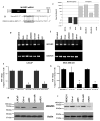
WAVE3 expression is elevated in cell lines with low levels of miR-31 microRNA. (A) Domain structure of the WAVE3 transcript showing the location of the seed sequence of miR-31 within the 3’UTR. (B) Alignment of the mature sequence of miR-31 shows a perfect match to a target sequence in the 3’UTR of WAVE3 cDNA, accession number NM_006646. (C). Inverse correlation between miR-31 and WAVE3 in invasive versus non-invasive cancer cell lines as determined by quantitative real-time RT-PCR. (D and E) Semi-quantitative RT-PCR analysis, (F and G) quantitative real-time RT-PCR analysis and (H and I) western blotting analysis of WAVE3 expression in LNCaP and MDA-MB-231 cells, respectively, transfected with miR-31 or siRNA against WAVE3. WAVE3 expression levels were markedly reduced in the miR-31- and siWAVE3-transfected cells. GAPDH was used as a control. *, p < 0.01 compared to untransfected or control-transfected cells.
Figure 2
Figure 2
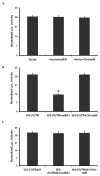
microRNA miR-31 directly targets the 3’UTR of WAVE3 and represses its expression. (A). Control experiment where firefly luciferase reporter plasmid pmirGlo control, was transiently transfected into MDA-MB-231 cells in the presence or absence of miR-31 or the control microRNA, and luciferase activities were measured after 48 h. (B) pmirGlo vector containing the entire 3’UTR of WAVE3 (W3-3’UTR), transiently transfected into MDA-MB-231 cells, in the presence or absence of miR-31 or the control microRNA, and luciferase activities were measured after 48 h. (C) pmirGlo vector containing WAVE3-3’UTR with deleted miR-31 seed sequence (W3-3’UTRd31), transiently transfected into MDA-MB-231 cells, in the presence or absence of miR-31 or the control microRNA, and luciferase activities were measured after 48 h. For all luciferase assays, Renilla luciferase activity was used for normalization. The data are the mean ± s. d. of at least 3 independent transfections. *, p < 0.01 compared to untransfected or control-transfected cells.
Figure 3
Figure 3
miR-31-mediated downregulation of WAVE3 expression inhibits cancer cell invasion in invasive cancer cells, while downregulation miR-31 increases invasion in non-invasive cancer cells. Quantification of invasive cells using a Matrigel invasion assay of MDA-MB-231 (A) and LNCaP (B) cells, transfected with either siRNA to WAVE3 or miR-31 precursor. (C) real-time quantitative RT-PCR and (D) Semi-quantitative RT-PCR quantification of WAVE3 in MCF7 cells transfected with either control microRNA or miR-31. (E) Quantification of invasive MCF7 cells transfected with either control microRNA or miR-31. Representative micrographs of invasive cells are shown in (F). At least 6 different fields were counted from each experiment. The data are the mean ± s. d. of at least 3 independent assays. *, p < 0.01 compared to untransfected or control-transfected cells.
Figure 4
Figure 4
Re-expression of miR200-resistant WAVE3 reverses miR31-mediated inhibition of LNCaP cell invasion. (A) Immunoblotting (IB) with the indicated antisera of MDA-MB-231 cells over-expressing GFP or GFP-WAVE3 fusion protein, and co-transfected with either siWAVE3 or miR-31. (B) IB with the indicated antisera of MDA-MB-231 cells over-expressing GFP or GFP-WAVE3 fusion protein, and co-transfected with either control siWAVE3 or control miR. (C and D) Quantification of invasive cells for the indicated treatment. At least 6 different fields were counted from each experiment. The data are the mean ± s. d. of at least 3 independent assays. *, p < 0.01 compared to untransfected or control-transfected cells.
Figure 5
Figure 5
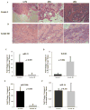
Expression of miR-31 correlates inversely with human breast cancer progression. (A) H&E staining of a representative Bloom-Richardson grade I invasive ductal carcinoma. At 1.5x, greater than 75% tubule formation is shown (150x original magnification). At 20x, the malignant glands are composed of uniform nuclei with fine chromatin (200x original magnification). At 40x, higher power detail of the uniform grade I nuclei that compose the carcinoma. The nuclei have fine chromatin, smooth nuclear membranes, and inconspicuous nucleoli. Mitotic figures are not readily identifiable (400x original magnification). (B) H&E staining of a representative Bloom-Richardson grade III invasive ductal carcinoma. At 1.5x, illustration of less than 10 tubules formed within this haphazard arrangement of malignant epithelium in a desmoplastic stroma (150x original magnification). At 20x, malignant cells display nuclear variability in size and shape and identifiable nucleoli (200x original magnification). At 40x, higher power detail of the grade III nuclei with marked variability in nuclear size and shape, nuclear membrane irregularities and readily identifiable nucleoli. Multiple mitotic figures are easily seen within this high-power field (400x original magnification). (C, D, E and F) Quantitative Real-time RT-PCR of miR-31, WAVE3, miR-200b and miR-16, respectively, in human primary Bloom-Richardson grade I and grade III invasive ductal carcinoma. The expression levels of each gene were normalized to GAPDH and plotted as fold change between the tumor and the corresponding normal tissue.
Figure 6
Figure 6
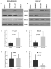
Expression of other breast cancer metastasis-relevant genes also correlates with breast cancer progression. (A and B) Western blotting analysis of WAVE3, RDX and ITGA5 in MDA-MB-231 (A), and LNCaP (B) with the indicated transient treatments. β-Actin was used as internal control for loading consistency. (C, D, E, and F) Quantitative Real-time RT-PCR of ITGA5, RhoA, RDX and E-Cad, respectively, in human primary Bloom-Richardson grade I and grade III invasive ductal carcinoma. The expression levels of each gene were normalized to GAPDH and plotted as fold change between the tumor and the corresponding normal tissue.
Figure 7
Figure 7
Mechanisms of WAVE3 regulation in cancer metastasis. Model illustrating the regulation of WAVE3 during the invasion-metastasis cascade. WAVE3 is regulated at the posttranscriptional level during the EMT process by miR-200, which results in increased cell motility, and. during the late stage of the invasion metastasis cascade, by miR-31 which results in increase in the invasive phenotype that is critical for tumor metastasis.

References

    1. Cory GO, Ridley AJ. Cell motility: braking WAVEs. Nature. 2002 Aug 15;418(6899):732–3. - PubMed
    1. Sossey-Alaoui K, Li X, Ranalli TA, Cowell JK. WAVE3-mediated cell migration and lamellipodia formation are regulated downstream of phosphatidylinositol 3-kinase. J Biol Chem. 2005 Jun 10;280(23):21748–55. - PubMed
    1. Sossey-Alaoui K, Ranalli TA, Li X, Bakin AV, Cowell JK. WAVE3 promotes cell motility and invasion through the regulation of MMP-1, MMP-3, and MMP-9 expression. Exp Cell Res. 2005 Aug 1;308(1):135–45. - PubMed
    1. Sossey-Alaoui K, Safina A, Li X, Vaughan MM, Hicks DG, Bakin AV, Cowell JK. Down-Regulation of WAVE3, a Metastasis Promoter Gene, Inhibits Invasion and Metastasis of Breast Cancer Cells. Am J Pathol. 2007 Jun;170(6):2112–21. - PMC - PubMed
    1. Takenawa T, Miki H. WASP and WAVE family proteins: key molecules for rapid rearrangement of cortical actin filaments and cell movement. J Cell Sci. 2001 May;114(Pt 10):1801–9. - PubMed

Publication types