

Technological advances in radiotherapy for esophageal cancer
- PMID: 21105188
- PMCID: PMC2992673
- DOI: 10.3748/wjg.v16.i44.5555
Technological advances in radiotherapy for esophageal cancer
Abstract
Radiotherapy with concurrent chemotherapy and surgery represent the main treatment modalities in esophageal cancer. The goal of modern radiotherapy approaches, based on recent technological advances, is to minimize post-treatment complications by improving the gross tumor volume definition (positron emission tomography-based planning), reducing interfraction motion (image-guided radiotherapy) and intrafraction motion (respiratory-gated radiotherapy), and by better dose delivery to the precisely defined planning target volume (intensity-modulated radiotherapy and proton therapy). Reduction of radiotherapy-related toxicity is fundamental to the improvement of clinical results in esophageal cancer, although the dose escalation concept is controversial.
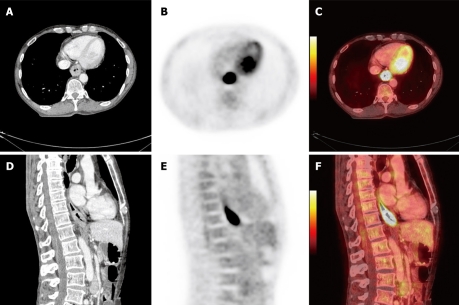
Figures

Similar articles
-
Tolerance and dose-volume relationship of intrathoracic stomach irradiation after esophagectomy for patients with thoracic esophageal squamous cell carcinoma.Oncotarget. 2015 Oct 13;6(31):32220-7. doi: 10.18632/oncotarget.4730. Oncotarget. 2015. PMID: 26314958 Free PMC article. Clinical Trial.
-
[Radiotherapy of the prostate gland--old wine in a new bottle?].Ther Umsch. 2006 Feb;63(2):151-6. doi: 10.1024/0040-5930.63.2.151. Ther Umsch. 2006. PMID: 16514968 Review. German.
-
Recent advances in radiotherapy treatment planning.Oncology (Williston Park). 1988 May;2(5):43-54, 57. Oncology (Williston Park). 1988. PMID: 3079328 Review.
-
[Radiotherapy in cancers of the oesophagus, the gastric cardia and the stomach].Cancer Radiother. 2016 Sep;20 Suppl:S161-8. doi: 10.1016/j.canrad.2016.07.039. Epub 2016 Aug 11. Cancer Radiother. 2016. PMID: 27523409 French.
-
Comparison of heart and coronary artery doses associated with intensity-modulated radiotherapy versus three-dimensional conformal radiotherapy for distal esophageal cancer.Int J Radiat Oncol Biol Phys. 2012 Aug 1;83(5):1580-6. doi: 10.1016/j.ijrobp.2011.10.053. Epub 2012 Jan 26. Int J Radiat Oncol Biol Phys. 2012. PMID: 22284687
Cited by
-
Survival benefit of perioperative chemotherapy for T1-3N0M0 stage esophageal cancer: a SEER database analysis.J Thorac Dis. 2021 Feb;13(2):995-1004. doi: 10.21037/jtd-20-2877. J Thorac Dis. 2021. PMID: 33717572 Free PMC article.
-
Intensity modulated radiotherapy (IMRT) with concurrent chemotherapy as definitive treatment of locally advanced esophageal cancer.Radiat Oncol. 2014 Aug 29;9:191. doi: 10.1186/1748-717X-9-191. Radiat Oncol. 2014. PMID: 25175056 Free PMC article. Clinical Trial.
-
A Meta-Analysis of Concurrent Chemoradiotherapy for Advanced Esophageal Cancer.PLoS One. 2015 Jun 5;10(6):e0128616. doi: 10.1371/journal.pone.0128616. eCollection 2015. PLoS One. 2015. PMID: 26046353 Free PMC article.
-
Correlation of CT texture changes with treatment response during radiation therapy for esophageal cancer: An exploratory study.PLoS One. 2019 Sep 26;14(9):e0223140. doi: 10.1371/journal.pone.0223140. eCollection 2019. PLoS One. 2019. PMID: 31557242 Free PMC article.
-
Retrospective Analysis of Dosimetric Comparison Between Intensity-Modulated Radiation Therapy and Volumetric-Modulated Arc Therapy in Patients With Esophageal Cancer.Cureus. 2025 Jan 5;17(1):e76981. doi: 10.7759/cureus.76981. eCollection 2025 Jan. Cureus. 2025. PMID: 39912037 Free PMC article.
References
-
- Koshy M, Esiashvilli N, Landry JC, Thomas CR Jr, Matthews RH. Multiple management modalities in esophageal cancer: epidemiology, presentation and progression, work-up, and surgical approaches. Oncologist. 2004;9:137–146. - PubMed
-
- Holmes RS, Vaughan TL. Epidemiology and pathogenesis of esophageal cancer. Semin Radiat Oncol. 2007;17:2–9. - PubMed
-
- Bosetti C, Levi F, Ferlay J, Garavello W, Lucchini F, Bertuccio P, Negri E, La Vecchia C. Trends in oesophageal cancer incidence and mortality in Europe. Int J Cancer. 2008;122:1118–1129. - PubMed
-
- Pera M, Manterola C, Vidal O, Grande L. Epidemiology of esophageal adenocarcinoma. J Surg Oncol. 2005;92:151–159. - PubMed
-
- National Comprehensive Cancer Network. Esophageal cancer. NCCN Clinical Practice Guidelines in Oncology. Version I. 2010. Accessed February 1. 2010. Available from: http://www.nccn.org/professionals/physician_gls/PDF/esophageal.pdf.
Publication types
MeSH terms
LinkOut - more resources
Full Text Sources
Medical
