Stability and membrane orientation of the fukutin transmembrane domain: a combined multiscale molecular dynamics and circular dichroism study
- PMID: 21105749
- PMCID: PMC3005826
- DOI: 10.1021/bi101743w
Stability and membrane orientation of the fukutin transmembrane domain: a combined multiscale molecular dynamics and circular dichroism study
Abstract
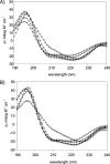
The N-terminal domain of fukutin-I has been implicated in the localization of the protein in the endoplasmic reticulum and Golgi Apparatus. It has been proposed to mediate this through its interaction with the thinner lipid bilayers found in these compartments. Here we have employed multiscale molecular dynamics simulations and circular dichroism spectroscopy to explore the structure, stability, and orientation of the short 36-residue N-terminus of fukutin-I (FK1TMD) in lipids with differing tail lengths. Our results show that FK1TMD adopts a stable helical conformation in phosphatidylcholine lipids when oriented with its principal axis perpendicular to the bilayer plane. The stability of the helix is largely insensitive to the lipid tail length, preventing hydrophobic mismatch by virtue of its mobility and ability to tilt within the lipid bilayers. This suggests that changes in FK1TMD tilt in response to bilayer properties may be implicated in the regulation of its trafficking. Coarse-grained simulations of the complex Golgi membrane suggest the N-terminal domain may induce the formation of microdomains in the surrounding membrane through its preferential interaction with 1,2-dipalmitoyl-sn-glycero-3-phosphatidylinositol 4,5-bisphosphate lipids.
Figures


Similar articles
-
Probing the oligomeric state and interaction surfaces of Fukutin-I in dilauroylphosphatidylcholine bilayers.Eur Biophys J. 2012 Feb;41(2):199-207. doi: 10.1007/s00249-011-0773-5. Epub 2011 Nov 11. Eur Biophys J. 2012. PMID: 22075563 Free PMC article.
-
Induction of nonbilayer structures in diacylphosphatidylcholine model membranes by transmembrane alpha-helical peptides: importance of hydrophobic mismatch and proposed role of tryptophans.Biochemistry. 1996 Jan 23;35(3):1037-45. doi: 10.1021/bi9519258. Biochemistry. 1996. PMID: 8547239
-
Influence of Lipid Saturation, Hydrophobic Length and Cholesterol on Double-Arginine-Containing Helical Peptides in Bilayer Membranes.Chembiochem. 2019 Nov 4;20(21):2784-2792. doi: 10.1002/cbic.201900282. Epub 2019 Sep 18. Chembiochem. 2019. PMID: 31150136 Free PMC article.
-
Hydrophobic mismatch between helices and lipid bilayers.Biophys J. 2003 Jan;84(1):379-85. doi: 10.1016/S0006-3495(03)74858-9. Biophys J. 2003. PMID: 12524291 Free PMC article.
-
Circular dichroism spectroscopy of membrane proteins.Chem Soc Rev. 2016 Sep 21;45(18):4859-72. doi: 10.1039/c5cs00084j. Epub 2016 Jun 27. Chem Soc Rev. 2016. PMID: 27347568 Review.
Cited by
-
The ELBA force field for coarse-grain modeling of lipid membranes.PLoS One. 2011;6(12):e28637. doi: 10.1371/journal.pone.0028637. Epub 2011 Dec 16. PLoS One. 2011. PMID: 22194874 Free PMC article.
-
Probing the oligomeric state and interaction surfaces of Fukutin-I in dilauroylphosphatidylcholine bilayers.Eur Biophys J. 2012 Feb;41(2):199-207. doi: 10.1007/s00249-011-0773-5. Epub 2011 Nov 11. Eur Biophys J. 2012. PMID: 22075563 Free PMC article.
-
Helical membrane protein conformations and their environment.Eur Biophys J. 2013 Oct;42(10):731-55. doi: 10.1007/s00249-013-0925-x. Epub 2013 Sep 1. Eur Biophys J. 2013. PMID: 23996195 Free PMC article. Review.
-
The NorM MATE transporter from N. gonorrhoeae: insights into drug and ion binding from atomistic molecular dynamics simulations.Biophys J. 2014 Jul 15;107(2):460-468. doi: 10.1016/j.bpj.2014.06.005. Biophys J. 2014. PMID: 25028887 Free PMC article.
-
Structural basis for plant plasma membrane protein dynamics and organization into functional nanodomains.Elife. 2017 Jul 31;6:e26404. doi: 10.7554/eLife.26404. Elife. 2017. PMID: 28758890 Free PMC article.
References
-
- Martin-Rendon E.; Blake D. J. (2003) Protein glycosylation in disease: New insights into the congenital muscular dystrophies. Trends Pharmacol. Sci. 24, 178–183. - PubMed
-
- Torelli S.; Brown S. C.; Brockington M.; Dolatshad N. F.; Jimenez C.; Skordis L.; Feng L. H.; Merlini L.; Jones D. H.; Romero N.; Wewer U.; Voit T.; Sewry C. A.; Noguchi S.; Nishino I.; Muntoni F. (2005) Sub-cellular localisation of fukutin related protein in different cell lines and in the muscle patients with MDC1C and LGMD2I. Neuromuscular Disord. 15, 836–843. - PubMed
-
- Keramaris-Vrantsis E.; Lu P. J.; Doran T.; Zillmer A.; Ashar J.; Esapa C. T.; Benson M. A.; Blake D. J.; Rosenfeld J.; Lu Q. L. (2007) Fukutin-related protein localizes to the Golgi apparatus and mutations lead to mislocalization in muscle in vivo. Muscle Nerve 36, 455–465. - PubMed
-
- Matsumoto H.; Noguchi S.; Sugie K.; Ogawa M.; Murayama K.; Hayashi Y. K.; Nishino I. (2004) Subcellular localization of fukutin and fukutin-related protein in muscle cells. J. Biochem. 135, 709–712. - PubMed
-
- Lommel M.; Willer T.; Strahl S. (2008) POMT2, a key enzyme in Walker-Warburg syndrome: Somatic sPOMT2, but not testis specific POMT2, is crucial for mannosyltransferase activity in vivo. Glycobiology 18, 615–625. - PubMed
Publication types
MeSH terms
Substances
Grants and funding
LinkOut - more resources
Full Text Sources
Molecular Biology Databases

