. 2011 Apr;117(2):197-208.
doi: 10.1111/j.1471-4159.2010.07119.x. Epub 2011 Mar 8.

Climbing fiber-evoked Purkinje cell discharge reduces expression of GABA(A) receptor-associated protein and decreases its interaction with GABA(A) receptors

Affiliations

Climbing fiber-evoked Purkinje cell discharge reduces expression of GABA(A) receptor-associated protein and decreases its interaction with GABA(A) receptors

Zuyuan Qian et al. J Neurochem. 2011 Apr.

Abstract

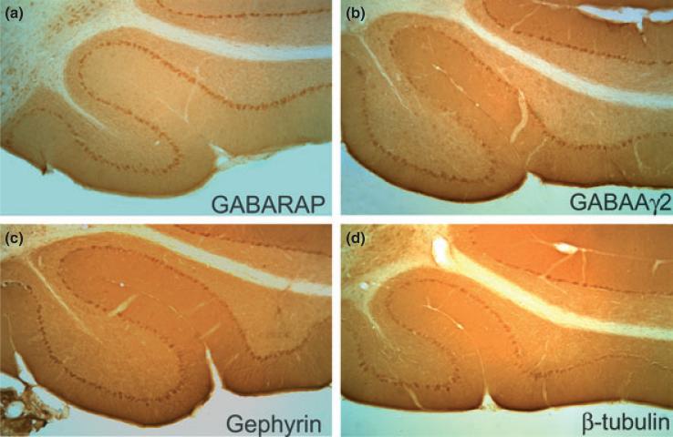
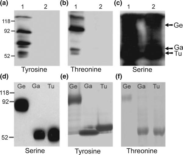
Sustained neuronal activity induces synaptic remodeling, in part, by altering gene expression. We have used a major climbing fiber pathway onto cerebellar Purkinje cells to investigate the effects of sustained climbing fiber-evoked glutamatergic synaptic transmission on transcription, expression and phosphorylation of proteins related to the regulation of inhibitory GABA(A) receptor function. Binocular horizontal optokinetic stimulation was used to modulate climbing fiber signals to Purkinje cells in the flocculus and nodulus of rabbits and mice. Purkinje cells in the flocculus and nodulus ipsilateral to the eye stimulated in the Posterior→Anterior direction received increased climbing fiber activity. Purkinje cells in flocculus and nodulus ipsilateral to the eye stimulated in the Anterior→Posterior direction received decreased climbing fiber activity. We identified changes in levels of gene transcripts in floccular and nodular Purkinje cells with the technique of differential display RT-PCR. Increased climbing fiber input reduced transcript levels and expression of GABA receptor-associated protein (GABARAP). Using a protein 'pull down' technique, we showed that GABARAP interacts with serine phosphorylated GABA(A)γ2, gephyrin and β-tubulin. Serine de-phosphorylation of GABA(A)γ2 reduced association of GABARAP with GABA(A)γ2. Climbing fiber activity did not influence the expression of GABA(A)γ2. Rather, it decreased its serine phosphorylation. Climbing fiber discharge decreased both expression of GABARAP and serine phosphorylation of GABA(A)γ2. Consequently, climbing fiber activity may reduce the surface expression of GABA(A) receptors in Purkinje cells rendering Purkinje cells less susceptible to interneuronal GABAergic inhibition.


Figures

Fig. 1
Fig. 1
Increased climbing fiber discharge decreases GABARAP transcripts in nodulus and flocculus. (a) Cartoon illustrates HOKS in the P → A direction with respect to the right eye. Horizontal direction-selective ganglion cells are excited by P → A stimulation and disfacilitated by A → P stimulation. Direction-selective ganglion cells project to the contralateral nucleus of the optic tract (NOT). NOT neurons project to neurons in the ipsilateral caudal dorsal cap (dc) of the inferior olive. Dorsal cap neurons project as climbing fibers to Purkinje cells in the contralateral flocculus (Fl). (b) In rabbits, binocular HOKS in P → A direction at 6 deg/s with respect to the left eye for 48 h increased climbing fiber activity in the left ventral nodulus and flocculus and reduced GABARAP transcripts in the left ventral nodulus and flocculus measured by Northern blot. (c) Decreased GABARAP transcripts in left nodulus and flocculus were confirmed by RT-PCR (24 cycles). GAPDH was used as a loading control for northern blot and RT-PCR. (d) Mice received either binocular or monocular HOKS in the P → A direction with respect to right eye for 24 h. An optical occluder prevented HOKS of the left eye in the A → P direction under the monocular condition. Both binocular and monocular HOKS reduced GABARAP transcripts in the right flocculus, detected by real-time PCR and normalized with respect to β-actin. Error bars indicate standard error of the mean. *p < 0.01, **p < 0.005, one tailed t-test.
Fig. 2
Fig. 2
HOKS-evoked climbing fiber activity modulates expression of GABARAP in nodulus and flocculus. Rabbits received HOKS for 48 h in the P → A direction with respect to left eye. (a) Western blots of protein extracts from the nodulus and flocculus show decreased expression of GABARAP in samples from the left nodulus and flocculus relative to those from the right. β-actin served as a loading control. (b) Histograms compare L/R ratio for GABARAP expression for five experiments and show that it is less than an L/R ratio = 1, indicating equal expression. Error bars indicate standard error of the mean. *p < 0.0001, one tailed t-test.
Fig. 3
Fig. 3
GABARAP associates with gephyrin, β-tubulin, GABAaγ2, PKC-γ and PKC-β in rabbit cerebellar lysates. Cerebellar lysates were co-incubated with his-tagged GABARAP. Pulled-down proteins were immunoprobed with antibodies to: (a) GABAaα1, (b) GABAaγ2, (c) gephyrin, (d) β-tubulin, (e) PKC-α, (f) PKC-β, (g) PKC-δ and (h) PKC-γ. Lane 1, cerebellar lysates. Lane 2, non-specific binding of the lysates to his-tagged beads was tested by co-incubating the lysates with his-tagged beads without GABARAP attached. Lane 3, specific binding was tested by co-incubating the lysates with his-tagged beads-attached to GABARAP. Lane 4, bacterially expressed, his-tagged GABARAP. Arrows indicate bands corresponding to targeted antigens in the lysates and in pulled down proteins.
Fig. 4
Fig. 4
Localization of GABARAP, GABAaγ2, gephyrin and β-tubulin to Purkinje cells. Sagittal sections from the mouse nodulus are immunolabeled with antibodies to: (a) GABARAP, (b) GABAaγ2, (c) gephyrin and (d) β-tubulin.
Fig. 5
Fig. 5
GABARAP associates with serine phosphorylated proteins in unstimulated cerebellar lysates. (a–c) Proteins from rabbit cerebellar lysates were pulled-down with his-tagged GABARAP and probed with antibodies to: (a) phosphotyrosine, (b) phosphothreonine and (c) phosphoserine. While all three phosphorylated proteins were found in cerebellar lysates, only serine-phosphorylated proteins were pulled down by GABARAP. (a–c) Lane 1 – proteins in cerebellar lysates, lane 2 – proteins pulled-down by his-tagged GABARAP. The arrows in (c) correspond to: gephyrin (Ge), GABAaγ2 (Ga) and β-tubulin (Tu). (d) The proteins pulled down by his-tagged GABARAP were immunoprecipitated with antibodies to GABAaγ2, gephyrin and β-tubulin and then immunoprobed with an antibody to phosphoserine, showing more clearly than in (c) that phosphorylated gephyrin, GABAaγ2 and β-tubulin are associated with GABARAP. (e,f) Antibodies to GABAaγ2, gephyrin and β-tubulin were used to immunoprecipitate proteins from cerebellar lysates. The immunoprecipitated proteins were probed with antibodies to: (e) phosphotyrosine and (f) phosphothreonine. The blots show that tyrosine- and threonine-phosphorylated gephyrin, GABAaγ2, and β-tubulin are expressed in the cerebellum, but do not associate with GABARAP.
Fig. 6
Fig. 6
Climbing fiber discharge does not modulate GABAa receptor subunit transcripts in mouse flocculus. (a) We examined the unstimulated cerebellum for transcription of 10 GABAa receptor subunit transcripts (α1–6, β1–3, γ2). Of these 10, six were transcribed (α1, α4, α6, β1, β2, γ2). (b) The density of RT-PCR bands in the left and right flocculi was measured for the six GABAa receptor subunits in a mouse that received HOKS for 24 h in the P → A direction with respect to the right eye. Cyclophilin mRNA (Cyclo) was co-transcribed and used as an equal loading control. (c) Histograms compare mean L/R ratios for the GABAa subunits for five mice following 24 h of HOKS. An L/R ratio = 1 indicates no differences in subunit transcription in the left and right flocculi. Error bars indicate standard error of the mean.
Fig. 7
Fig. 7
Serine phosphorylation of GABAaγ2 is reduced in vivo by climbing fiber discharge and increased in vitro by treatment with PMA. (a) Following 48 h of HOKS, protein extracts were prepared from the left and right nodulus of rabbits and immunoprecipitated with antibodies to: (a-i) Gephyrin, (a-ii) GABAaγ2 and (a-iii) β-tubulin. Equal levels of immunoprecipitates were confirmed for each lane by immunoblotting with antibodies to gephyrin, GABAaγ2 and β-tubulin. Blots were stripped and re-probed with an antibody to phosphoserine. (a-iv) Histograms confirm that increased climbing fiber activity in the left nodulus decreased serine phosphorylation of GABAaγ2, but not of gephyrin or β-tubulin. (b) In vitro PMA treatment of cerebellar slices increased serine phosphorylation of gephyrin, GABAaγ2 and β-tubulin. Lysates were prepared from nodular slices previously incubated for 1 h with: (i) untreated control, (ii) αPMA at 250 nM and (iii) PMA at 250 nM. Proteins from the lysates were immunoprecipitated with antibodies to: (b-i) gephyrin, (b-ii) GABAaγ2 and (b-iii) β-tubulin. The immunoprecipitates were probed with an antibody to phosphoserine. (b-iv) Histograms show that PMA treatment increased serine phosphorylation of all three proteins relative to the αPMA controls. (c) GABARAP immunoprecipitated serine phosphorylated gephyrin and GABAaγ2 after incubation in vitro in PMA. Cerebellar slices were incubated for 1 h with: (i) untreated control, (ii) αPMA at 250 nM or (iii) PMA at 250 nM. Lysate proteins were immunoprecipitated with an antibody to GABARAP. Immunoprecipitates were probed with antibodies to: (c-i) gephyrin, (c-ii) GABAaγ2 and (c-iii) β-tubulin. (c-iv) Histograms show that more GABAaγ2 and gephyrin co-immunopreciptiated with GABARAP in PMA treated slices than in αPMA-treated controls. Error bars indicate standard error of the mean. Asterisks indicate statistical significance of Student's t-test, p < 0.05).
Fig. 8
Fig. 8
Dephosphorylation of GABAaγ2 reduces its association with GABARAP. (a) GABAaγ2 was immunoprecipitated from rabbit cerebellar lysates (lanes 1 and 2). (b) One of the immunoprecipitates of GABAaγ2 was dephosphorylated (lane 2) by treatment with PP1. The untreated control GABAaγ2 (lane 1) and the PP1 treated GABAaγ2 were probed with an antibody to phosphoserine, confirming reduced serine phosphorylation in the PP1 treated GABAaγ2. (c) Following PP1 treatment of GABAaγ2, histidine-tagged GABARAP was used to pull down associated proteins. The pulled down proteins were probed with an antibody to GABAaγ2. Histidine-tagged GABARAP pulled down less GABAaγ2 in the PP1 treated sample (upper bands, lane 2). Lower bands indicate equal amounts of GABARAP used to pull down GABAaγ2 in the control and PP1 treated samples.
Fig. 9
Fig. 9
In vivo and in vitro regulation of GABAa receptors on Purkinje cells. (a) Increased climbing fiber discharge evoked by HOKS in vivo reduced: (1) Expression of GABARAP (orange pentagon) and (2) Serine-phosphorylation of GABAaγ2. Both effects could reduce GABAa receptor clustering in Purkinje cell cytoplasmic membrane. (b) In vitro incubation of cerebellar slices with PMA activates PKC. This enhances serine phosphorylation of: (1) GABAaγ2, (2) β-tubulin and (3) gephyrin. Serine phosphorylation increased the association of these proteins with GABARAP and could influence other key components responsible for formation, insertion and clustering of GABAa receptors in Purkinje cell cytoplasmic membrane. NSF, N-ethylmaleimide-sensitive factor; Ps, phosphoserine residue.

Similar articles

Cited by

References

    1. Arancibia-Cárcamo IL, Kittler JT. Regulation of GABA(A) receptor membrane trafficking and synaptic localization. Pharmacol. Ther. 2009;123:17–31. - PubMed
    1. Barmack NH, Nelson BJ. Influence of long-term optokinetic stimulation on eye movements of the rabbit. Brain Res. 1987;437:111–120. - PubMed
    1. Barmack NH, Qian Z. Activity-dependent expression of calbindin in rabbit floccular Purkinje cells modulated by opto-kinetic stimulation. Neuroscience. 2002;113:235–250. - PubMed
    1. Barmack NH, Shojaku H. Vestibular and visual signals evoked in the uvula-nodulus of the rabbit cerebellum by natural stimulation. J. Neurophysiol. 1995;74:2573–2589. - PubMed
    1. Barmack NH, Yakhnitsa V. Cerebellar climbing fibers modulate simple spikes in cerebellar Purkinje cells. J. Neurosci. 2003;23:7904–7916. - PMC - PubMed

Publication types

MeSH terms