. 2010 Nov 24:10:81.
doi: 10.1186/1472-6750-10-81.

Improved Tet-responsive promoters with minimized background expression

Affiliations

Improved Tet-responsive promoters with minimized background expression

Rainer Loew et al. BMC Biotechnol. .

Abstract

Background: The performance of the tetracycline controlled transcriptional activation system (Tet system) depends critically on the choice of minimal promoters. They are indispensable to warrant low expression levels with the system turned "off". On the other hand, they must support high level of gene expression in the "on"-state.

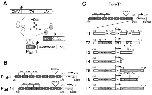
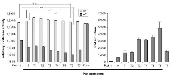
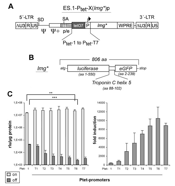
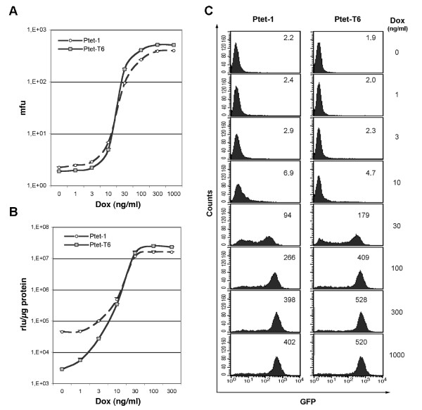
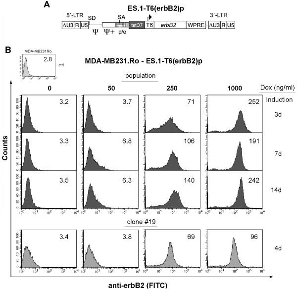
Results: In this study, we systematically modified the widely used Cytomegalovirus (CMV) minimal promoter to further minimize background expression, resulting in an improved dynamic expression range. Using both plasmid-based and retroviral gene delivery, our analysis revealed that especially background expression levels could be significantly reduced when compared to previously established "standard" promoter designs. Our results also demonstrate the possibility to fine-tune expression levels in non-clonal cell populations. They also imply differences regarding the requirements for tight regulation and high level induction between transient and stable gene transfer systems.

Conclusions: Until now, our understanding of mammalian transcriptional regulation including promoter architecture is limited. Nevertheless, the partly empirical modification of cis-elements as shown in this study can lead to the specific improvement of the performance of minimal promoters. The novel composite Ptet promoters introduced here will further expand the utility of the Tet system.


Figures

Figure 1
Figure 1
Outline of the Tet System and the Ptet promoters. (A) The tet-responsive transactivator (here tTA, i.e. Tet-Off system) is constitutively expressed in the cell. tTA homodimers bind to the heptameric tet operator sequence (tetO7) in the absence of Dox (a tetracycline derivative). They dissociate from their recognition sites after Dox addition. The Dox response would be reversed for "Tet-On" type transactivator lines also used in this study (C = CMV minimal promoter). (B) Comparison of Ptet-1, the originally described Ptet promoter and the commercially available Ptet-14 ("Ptight"). Both consist of tet operator heptamers created by monomer ligation via compatible restriction sites (Xho/Sal), but with differently spaced operator centers. The CMV-minimal promoters differ in size, but both contain the authentic TATA-box and initiator sequence (Inr). (C) The newly designed Ptet promoters are shown, consisting of a tet operator heptamer with 36nt spacing, and randomized fusion points between all the operators. The CMV derived minimal promoters of the T2-T7 Ptet promoters all have a consensus ("c") TATA-box and TFIIB binding site but differ in the composition of the 5' UTR as outlined in the text. Point mutations are indicated by (*), deletions by dashed lines. Nucleotide positions are given relative to the transcriptional start site (+1). The initiator (Inr) and downstream promoter element (DPE) is indicated.
Figure 2
Figure 2
Transient transfection analysis of the new Ptet promoters in HeLa-EM2 cells. Left panel: Firefly luciferase activities after transfection of HeLa-EM2 cells with the new Ptet promoter driven reporter constructs were determined in the on-(+Dox) and off-state (-Dox) of the system. Values were normalized to the activity of a cotransfected internal standard (beta-galactosidase reporter). Statistical comparison of Ptet-1 and Ptet-14 with Ptet-T6 via students t-test was performed with Graph Pad Prism version 5.03 software (** = p-value < 0,01, *** = p-value < 0.001, n.s. = not significant). High amounts of luciferase reporter plasmids were transfected to achieve relative light unit readings above instrument background that could be reliably quantified. For example, in this experiment the instrument background (identical to mock transfected cells) was 1,5 × 10² rlu while that of extracts from Ptet-T6 transfected cells (-Dox) was 3,5 × 10² rlu. Right panel: Regulation factors for the individual Ptet promoters from the analysis shown left. The results shown are the mean of three independent transfection experiments, the error bars represent the SEM.
Figure 3
Figure 3
Retroviral transfer of the new Ptet promoters. (A) The different regulatory units were inserted into the γ-retroviral self-inactivating (SIN) vector „ES.1". The U3-enhancer elements (ΔU3) were deleted from the provirus, while the enlarged packaging region (ψ,ψ+) as well as the native splice acceptor (SA) located in the pol/env region of the virus were retained. In order to enhance viral titers and translational efficiency, a woodchuck posttranscriptional regulatory element (WPRE) was integrated 3'-to the reporter gene. (B) The lmg* dual reporter was used throughout all experiments with the viral vectors. The corresponding gene consists of the firefly luciferase open reading frame (orf), with deleted stop codon, the 3'-half of the troponin C α-helix5 and the eGFP orf with the deleted start codon. (C) Left panel: Determination of specific luciferase activity (relative light units, rlu) that were obtained from transduced, FACS enriched HtTA-1 cell populations. Statistical comparison was done as described in figure 2. Right panel: Regulation factors for the individual Ptet promoters from the analysis shown left. The results shown are derived from at least two cell populations generated independently. Each population was analyzed two to three times; the error bars represent the SEM.
Figure 4
Figure 4
Dose response analyses of Ptet-1 and Ptet-T6 in transduced HeLa-EM2 cell populations. (A) Determination of dose response via eGFP function of lmg*. Mean fluorescence units (mfu) are given for the complete populations (≥ 95% purity). For value ranges see insets in "C". (B) The simultaneous analysis of identical populations for specific luciferase activity of lmg*. The rlu values ranged from about 4.5 × 10⁴ (± 2.8 × 10³) to 1.6 × 10⁷ (± 4.7 × 10⁵) rlu/μg for Ptet-1 and 2.9 × 10³ (± 1.1 × 10²) to 2.3 × 10⁷ (± 3.9 × 10⁵) rlu/μg for Ptet-T6 in the off-and on-state of the system. (C) FACS-analysis of a representative cell population for Ptet-1 and Ptet-T6 promoters transferred by the viral vectors. Mfu values of the whole population were given as inset. The cellular background of the GFP-negative Hela-EM2 parental cell line was 1.83. The Dox concentrations used are indicated. Induction was for 96 hours. Results shown in figs. A and B were obtained from two independently generated populations for each vector.
Figure 5
Figure 5
Transduction of a Ptet-T6 regulated erbB2 expression unit into MDA-MB231.Ro cells. (A) γ-retroviral SIN-vector used to transfer erbB2 into rtTA-S2-positive MDA-MB231.Ro target cells. (B) FACS-analysis of transduced cell populations induced for the indicated time and Dox concentrations. The expression of the erbB2 transgene was detected by a monoclonal antibody (mAB ALX-804-573; Alexis) in combination with a fluorescence labeled secondary antibody (donkey anti mouse IgG-FITC, affipure, Jackson Immuno Res.). Mean fluorescence units (mfu, blot inset) are given for the complete population (≥ 92% purity). Clone #19 was derived from this population by limiting dilution.

Similar articles

Cited by

References

    1. Gossen M, Bujard H. Tight control of gene expression in mammalian cells by tetracycline-responsive promoters. Proc Natl Acad Sci USA. 1992;89(12):5547–5551. doi: 10.1073/pnas.89.12.5547. - DOI - PMC - PubMed
    1. Gossen M, Freundlieb S, Bender G, Muller G, Hillen W, Bujard H. Transcriptional activation by tetracyclines in mammalian cells. Science. 1995;268(5218):1766–1769. doi: 10.1126/science.7792603. - DOI - PubMed
    1. Park HJ, RajBhandary UL. Tetracycline-regulated suppression of amber codons in mammalian cells. Mol Cell Biol. 1998;18(8):4418–4425. - PMC - PubMed
    1. Yoshida Y, Hamada H. Adenovirus-mediated inducible gene expression through tetracycline-controllable transactivator with nuclear localization signal. Biochem Biophys Res Commun. 1997;230(2):426–430. doi: 10.1006/bbrc.1996.5975. - DOI - PubMed
    1. Wells KD, Foster JA, Moore K, Pursel VG, Wall RJ. Codon optimization, genetic insulation, and an rtTA reporter improve performance of the tetracycline switch. Transgenic Res. 1999;8(5):371–381. doi: 10.1023/A:1008952302539. - DOI - PubMed

Publication types

LinkOut - more resources