

Novel insights into the function of β-cell M3 muscarinic acetylcholine receptors: therapeutic implications
- PMID: 21106385
- PMCID: PMC3053051
- DOI: 10.1016/j.tem.2010.10.004
Novel insights into the function of β-cell M3 muscarinic acetylcholine receptors: therapeutic implications
Abstract
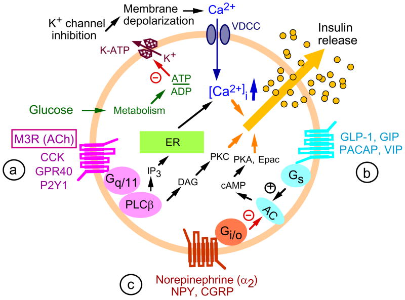
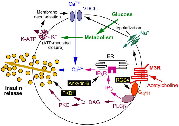
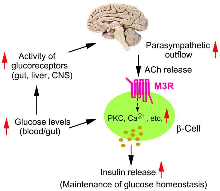
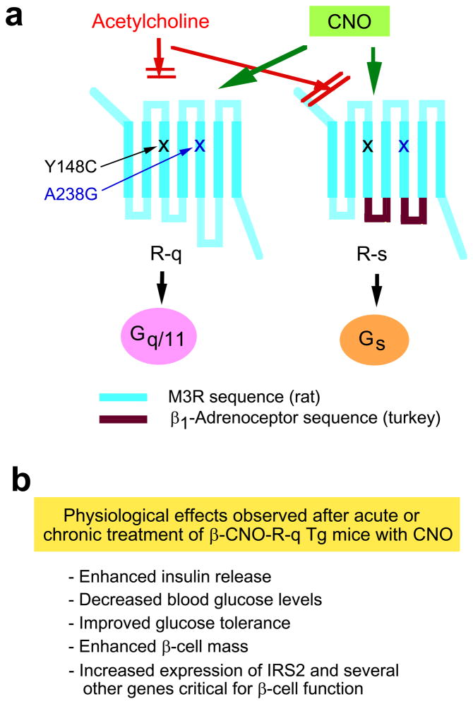
Impaired function of pancreatic β-cells is one of the hallmarks of type 2 diabetes. β-cell function is regulated by the activity of many hormones and neurotransmitters, which bind to specific cell surface receptors. The M(3) muscarinic acetylcholine receptor (M3R) belongs to the superfamily of G protein-coupled receptors and, following ligand dependent activation, selectively activates G proteins of the G(q/11) family. Recent studies with M3R mutant mice strongly suggest that β-cell M3Rs play a central role in promoting insulin release and maintaining correct glucose homeostasis. In this review, we highlight recent studies indicating that β-cell M3Rs and components of downstream signaling pathways might represent promising new targets for the treatment of type 2 diabetes.
Published by Elsevier Ltd.
Figures
Similar articles
-
Allosteric modulation of β-cell M3 muscarinic acetylcholine receptors greatly improves glucose homeostasis in lean and obese mice.Proc Natl Acad Sci U S A. 2019 Sep 10;116(37):18684-18690. doi: 10.1073/pnas.1904943116. Epub 2019 Aug 26. Proc Natl Acad Sci U S A. 2019. PMID: 31451647 Free PMC article.
-
Minireview: Novel aspects of M3 muscarinic receptor signaling in pancreatic β-cells.Mol Endocrinol. 2013 Aug;27(8):1208-16. doi: 10.1210/me.2013-1084. Epub 2013 Jul 2. Mol Endocrinol. 2013. PMID: 23820900 Free PMC article. Review.
-
Beneficial metabolic effects caused by persistent activation of beta-cell M3 muscarinic acetylcholine receptors in transgenic mice.Endocrinology. 2010 Nov;151(11):5185-94. doi: 10.1210/en.2010-0519. Epub 2010 Sep 15. Endocrinology. 2010. PMID: 20843999 Free PMC article.
-
A critical role for beta cell M3 muscarinic acetylcholine receptors in regulating insulin release and blood glucose homeostasis in vivo.Cell Metab. 2006 Jun;3(6):449-61. doi: 10.1016/j.cmet.2006.04.009. Cell Metab. 2006. PMID: 16753580
-
Metabolic roles of the M3 muscarinic acetylcholine receptor studied with M3 receptor mutant mice: a review.J Recept Signal Transduct Res. 2008;28(1-2):93-108. doi: 10.1080/10799890801942002. J Recept Signal Transduct Res. 2008. PMID: 18437633 Review.
Cited by
-
Vagal nerve stimulation improves mitochondrial dynamics via an M3 receptor/CaMKKβ/AMPK pathway in isoproterenol-induced myocardial ischaemia.J Cell Mol Med. 2017 Jan;21(1):58-71. doi: 10.1111/jcmm.12938. Epub 2016 Aug 5. J Cell Mol Med. 2017. PMID: 27491814 Free PMC article.
-
Neurofunctional imaging of β-cell dynamics.Diabetes Obes Metab. 2012 Oct;14 Suppl 3(0 3):91-100. doi: 10.1111/j.1463-1326.2012.01651.x. Diabetes Obes Metab. 2012. PMID: 22928569 Free PMC article. Review.
-
Loss of Free Fatty Acid Receptor 2 leads to impaired islet mass and beta cell survival.Sci Rep. 2016 Jun 21;6:28159. doi: 10.1038/srep28159. Sci Rep. 2016. PMID: 27324831 Free PMC article.
-
Allosteric modulation of β-cell M3 muscarinic acetylcholine receptors greatly improves glucose homeostasis in lean and obese mice.Proc Natl Acad Sci U S A. 2019 Sep 10;116(37):18684-18690. doi: 10.1073/pnas.1904943116. Epub 2019 Aug 26. Proc Natl Acad Sci U S A. 2019. PMID: 31451647 Free PMC article.
-
Loss of FFA2 and FFA3 increases insulin secretion and improves glucose tolerance in type 2 diabetes.Nat Med. 2015 Feb;21(2):173-7. doi: 10.1038/nm.3779. Epub 2015 Jan 12. Nat Med. 2015. PMID: 25581519
References
-
- Kahn SE. The relative contributions of insulin resistance and beta-cell dysfunction to the pathophysiology of Type 2 diabetes. Diabetologia. 2003;46:3–19. - PubMed
-
- Ahrén B. Islet G protein-coupled receptors as potential targets for treatment of type 2 diabetes. Nat Rev Drug Discov. 2009;8:369–385. - PubMed
-
- Baggio LL, Drucker DJ. Biology of incretins: GLP-1 and GIP. Gastroenterology. 2007;132:2131–2157. - PubMed
Publication types
MeSH terms
Substances
Grants and funding
LinkOut - more resources
Full Text Sources
