. 2011 Feb 18;286(7):5034-42.
doi: 10.1074/jbc.M110.155903. Epub 2010 Nov 24.

The tumor suppressor hTid1 inhibits STAT5b activity via functional interaction

Affiliations

The tumor suppressor hTid1 inhibits STAT5b activity via functional interaction

Isabelle Dhennin-Duthille et al. J Biol Chem. .

Abstract

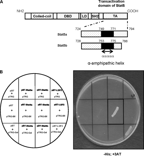
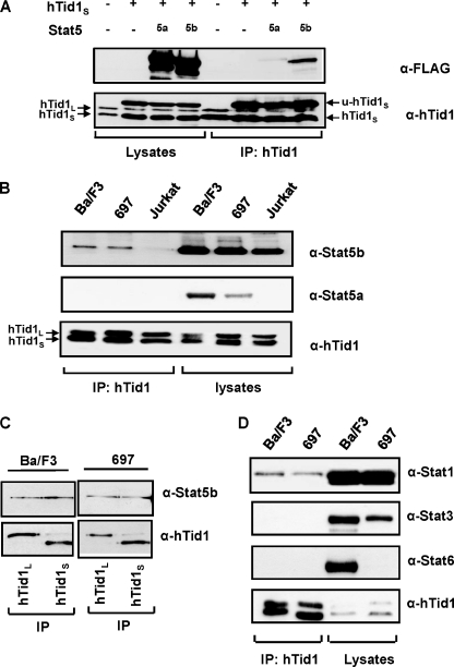
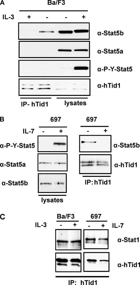
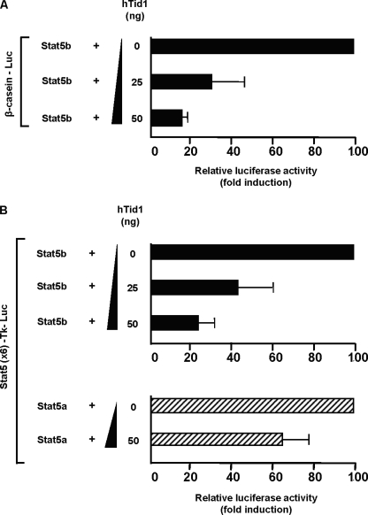
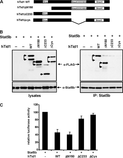
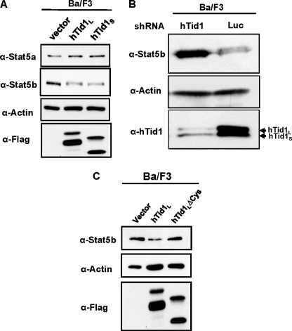
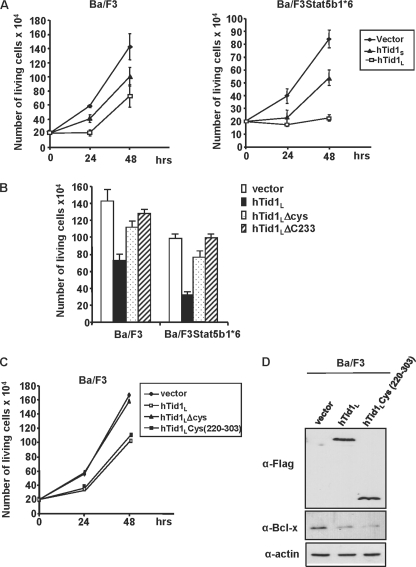
STAT5a and -5b (signal transducers and activators of transcription 5a and 5b) proteins play an essential role in hematopoietic cell proliferation and survival and are frequently constitutively active in hematologic neoplasms and solid tumors. Because STAT5a and STAT5b differ mainly in the carboxyl-terminal transactivation domain, we sought to identify new proteins that bind specifically to this domain by using a bacterial two-hybrid screening. We isolated hTid1, a human DnaJ protein that acts as a tumor suppressor in various solid tumors. hTid1 interacts specifically with STAT5b but not with STAT5a in hematopoietic cell lines. This interaction involves the cysteine-rich region of the hTid1 DnaJ domain. We also demonstrated that hTid1 negatively regulates the expression and transcriptional activity of STAT5b and suppresses the growth of hematopoietic cells transformed by an oncogenic form of STAT5b. Our findings define hTid1 as a novel partner and negative regulator of STAT5b.


Figures

FIGURE 1.
FIGURE 1.
The transactivation domain of STAT5b interacts with hTid1 in bacterias. A, schematic representation of the transactivation domain (TA) of STAT5a and STAT5b used in the bacterial two-hybrid screening. DBD, DNA-binding domain; SH2, Src homology 2 domain. B, bacterias were transformed with the plasmid pTRG-B6 (containing the DnaJ domain of hTid-1) and pBT-STAT5a, pBT-STAT5b, or the empty vector pBT as indicated. Bacterias were also transformed with the constructs pBT-LGF2 and pTRG-Gal11P as positive controls. The transformed cells were selected on plates containing the inhibitor 3-amino-1,2,4-triazole (+3AT) and lacking the amino acid histidine. The presence of transformed bacterias on the plate containing selective medium is shown. LD, Linker domain.
FIGURE 2.
FIGURE 2.
Association of STAT5b and hTid1 in transfected cells and hematopoietic cell lines. A, transfected COS cell extracts expressing FLAG-tagged STAT5a or FLAG-tagged STAT5b and the short form of hTid1 (hTid1S) were immunoprecipitated (IP) with an anti-hTid1 antibody. The association of STAT5a and STAT5b with hTid1S was then analyzed by Western blot using an anti-FLAG antibody. Endogenous hTid1L and hTid1S proteins are shown on the left, whereas the two forms of hTid1S (the unprocessed and mature forms, u-hTid1S and hTid1S) are shown on the right. B, STAT5b interacts with hTid1 in various murine and human lymphoid cell lines. Ba/F3, Jurkat, and 697 cell extracts were immunoprecipitated with an anti-hTid1 antibody, and the presence of STAT5 in the immunoprecipitates was detected by Western blot using anti-STAT5a and anti-STAT5b antibodies. C, STAT5b interacts with the long form (hTid1L) or the short form (hTid1S) of hTid1. Ba/F3 and 697 cell extracts were immunoprecipitated with antibodies that specifically recognize hTid1L or hTid1S. The presence of STAT5b in the immunoprecipitates was then determined by Western blot using STAT5b antibodies. D, interaction of STAT1 but not STAT3 or STAT6 with hTid1. Ba/F3 and 697 cell extracts were immunoprecipitated with an anti-hTid1 antibody. The presence of STAT1, STAT3, or STAT6 in the immunoprecipitates was then determined by Western blot using the indicated antibodies.
FIGURE 3.
FIGURE 3.
Dissociation of the STAT5b-hTid1 complex upon ligand-induced tyrosine phosphorylation of STAT5. A, extracts from Ba/F3 cells treated or not with IL-3 for 30 min were immunoprecipitated (IP) with an anti-hTid1 antibody, and the presence of STAT5 was determined by Western blot analysis with the indicated antibodies. B, a similar experiment was conducted with the human 697 pre-B cells treated with IL-7. C, the association of STAT1 with hTid1 was also analyzed by Western blot after immunoprecipitation of hTid1 from unstimulated or stimulated Ba/F3 and 697 cell extracts.
FIGURE 4.
FIGURE 4.
hTid1 suppresses the transcriptional activity of STAT5b. A, COS cells were transfected with the STAT5-specific β-casein promoter luciferase construct (20 ng), the STAT5b (25 ng), and the prolactin receptor (30 ng) expression vectors and increasing amounts of hTid1S plasmid DNA as indicated. Transfected COS cells were stimulated with prolactin (5 ng/ml) and lysed, and the luciferase activities were determined. Results are the mean of four independent experiments. B, a similar experiment was conducted with a thymidine kinase-luciferase reporter construct containing six copies of the STAT5 response element of the β-casein promoter (Stat5 × 6-Tk-luciferase). The specific inhibition of STAT5b transcriptional activity induced by hTid1 was also evaluated in cells transfected with a STAT5a expression vector. Error bars, S.D.
FIGURE 5.
FIGURE 5.
hTid1 interacts with and inhibits STAT5b transcriptional activity via the central DnaJ cysteine-rich region. A, schematic representation of the hTid1 mutants. The position of the different DnaJ domains is shown. B, FLAG-tagged WT hTid1 and deletion mutants of hTid1 were transfected with a STAT5b expression vector in COS cells. Expression of the different hTid1 constructs and STAT5b was first verified by Western blot with an anti-FLAG antibody or an anti-STAT5b antibody (left). STAT5b was then immunoprecipitated (IP), and the presence of the different hTid1 variants in the immunoprecipitates was detected by Western blot with an anti-FLAG antibody. Membrane was reprobed with an anti-STAT5b antibody. The two arrows indicate the mature and non processed forms that were detected following transfection with the different hTid1 constructs. C, the inhibitory effect of the hTid1 mutants on the transcriptional activity of STAT5b was next evaluated in transfected COS cells by using the STAT5 reporter assay described in the legend to Fig. 3B. Results are the mean of four independent experiments. Error bars, S.D.
FIGURE 6.
FIGURE 6.
hTid1 inhibits STAT5b but not STAT5a expression in Ba/F3 cells. A, Ba/F3 cells were electroporated with the empty vector or the FLAG-tagged hTid1L and hTid1S constructs as indicated. The following day, GFP+ cells were sorted, and the levels of STAT5a and STAT5b were next determined by immunoblotting with specific antibodies. Membranes were reprobed with an anti-actin antibody. Expression of hTid1L and hTid1S was also verified by Western blot with an anti-FLAG antibody (the two bands represent the mature and unprocessed forms of the FLAG-tagged hTid1L or hTid1S protein). B, Ba/F3 cells were electroporated with the psiRNAhTid1-h7SKGFP plasmid or the psiRNA-luc-h7SKGFP construct as control. GFP+ cells were sorted by flow cytometry, and expression levels of hTid1 and STAT5b were then analyzed by Western blot using the indicated antibodies. The membrane was reprobed with an anti-actin antibody. C, hTid1L and hTid1LΔCys constructs were transfected in Ba/F3 cells. After cell sorting, expression of STAT5b was determined by Western blot using an anti-STAT5b antibody. Levels of hTid1L and hTid1LΔCys proteins were also determined by Western blot with an anti-FLAG antibody (the two bands represent the mature and unprocessed forms of the FLAG-tagged hTid1L or hTid1LΔCys protein). Membrane was reprobed with an anti-actin antibody.
FIGURE 7.
FIGURE 7.
The DnaJ cysteine-rich domain of hTid1 is required for inhibition of cell growth and STAT5 activity in hematopoietic cells. A, parental Ba/F3 cells and Ba/F3 cells transformed by the constitutively active STAT5b1*6 mutant (Ba/F3STAT5b1*6) were electroporated with the FLAG-tagged hTid1L and hTid1S constructs or the empty vector as indicated. GFP+ cells were sorted 24 h later, and the number of living cells was daily enumerated. Results are the mean of three independent experiments. B, cells were also transfected with hTid1 mutants lacking the DnaJ cysteine-rich region (hTid1ΔCys and hTid1Δ233). Growth rates of transfected cells were determined 48 h after cell sorting. Results are the mean of three independent experiments. C, the DnaJ cysteine-rich domain of hTid1 is sufficient to block the growth of Ba/F3 cells. Ba/F3 cells were transfected with the hTid1 or hTid1ΔCys constructs or a vector expressing the FLAG-tagged DnaJ cysteine-rich domain of hTid1 (hTid1Lcys(220–303)). GFP+ cells were sorted 24 h later, and living cells were counted daily. Results are the mean of three independent experiments. D, the DnaJ cysteine-rich domain of hTid1 is sufficient to inhibit Bcl-xL expression in Ba/F3 cells. Cell extracts from GFP+ Ba/F3 cells transfected with the wild-type hTid1L or the hTid1 mutant hTid1Lcys(220–303) construct were analyzed by Western blot using anti-Bcl-xL antibody. Levels of hTid1L and hTid1LΔCys proteins were also determined by Western blot with an anti-FLAG antibody. Membrane was reprobed with an anti-actin antibody.

Similar articles

Cited by

References

    1. Leonard W. J. (2001) Int. J. Hematol. 73, 271–277 - PubMed
    1. Liu X., Robinson G. W., Gouilleux F., Groner B., Hennighausen L. (1995) Proc. Natl. Acad. Sci. U.S.A. 92, 8831–8835 - PMC - PubMed
    1. Buitenhuis M., Coffer P. J., Koenderman L. (2004) Int. J. Biochem. Cell Biol. 36, 2120–2124 - PubMed
    1. Liu X., Robinson G. W., Wagner K. U., Garrett L., Wynshaw-Boris A., Hennighausen L. (1997) Genes Dev. 11, 179–186 - PubMed
    1. Udy G. B., Towers R. P., Snell R. G., Wilkins R. J., Park S. H., Ram P. A., Waxman D. J., Davey H. W. (1997) Proc. Natl. Acad. Sci. U.S.A. 94, 7239–7244 - PMC - PubMed

Publication types

MeSH terms