. 2011 Feb 25;286(8):6641-9.
doi: 10.1074/jbc.M110.168872. Epub 2010 Nov 24.

Post-transcriptional defects of antioxidant selenoenzymes cause oxidative stress under methylmercury exposure

Affiliations

Post-transcriptional defects of antioxidant selenoenzymes cause oxidative stress under methylmercury exposure

Fusako Usuki et al. J Biol Chem. .

Abstract

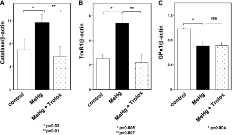
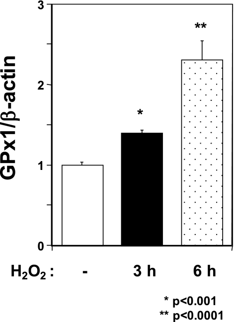
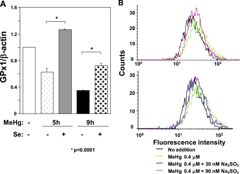
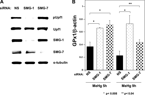
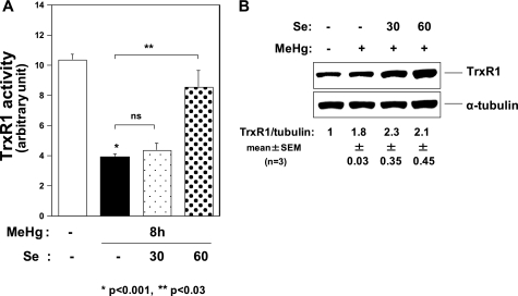
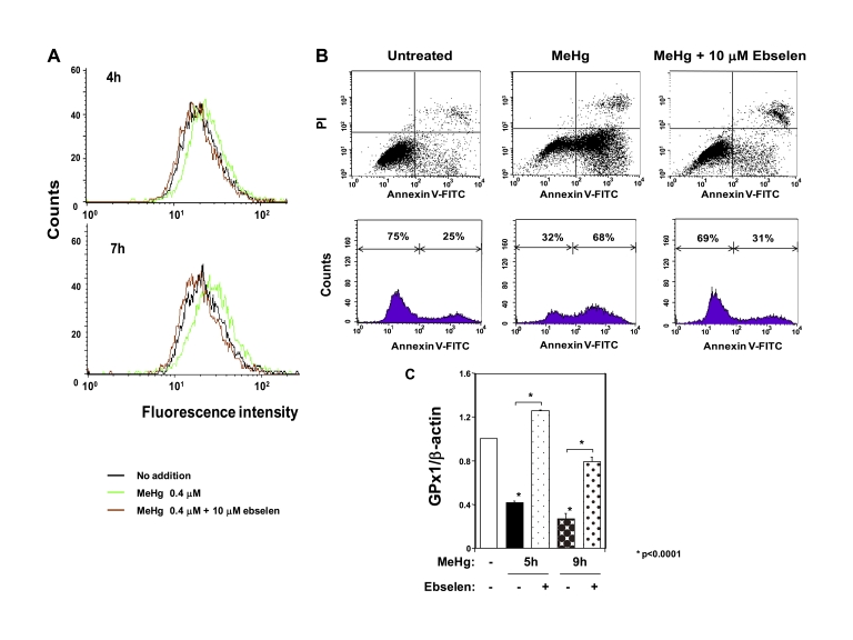
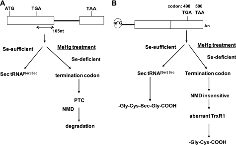
Methylmercury (MeHg) toxicity is a continuous environmental problem to human health. The critical role of oxidative stress in the pathogenesis of MeHg cytotoxicity has been clarified, but the molecular mechanisms underlying MeHg-mediated oxidative stress remain to be elucidated. Here we demonstrate a post-transcriptional effect of MeHg on antioxidant selenoenzymes by using a MeHg-susceptible cell line. MeHg-induced selenium deficiency leads to failure of the recoding of a UGA codon for selenocysteine and results in degradation of the major antioxidant selenoenzyme glutathione peroxidase 1 (GPx1) mRNA by nonsense-mediated mRNA decay (NMD), a cellular mechanism that detects the premature termination codon (PTC) located 5'-upstream of the last exon-exon junction and degrades PTC-containing mRNAs. In contrast, thioredoxin reductase 1 (TrxR1), another antioxidant selenoenzyme of the thioredoxin system, was likely skipped by NMD because of a UGA codon in the last exon. However, TrxR1 activity was decreased despite mRNA up-regulation, which was probably due to the synthesis of aberrant TrxR1 protein without selenocysteine. Changes in selenoenzyme GPx1 and TrxR1 mRNAs were observed earlier than was the incidence of oxidative stress and up-regulation of other antioxidant enzyme mRNAs. Results indicated that the MeHg-induced relative selenium-deficient condition affects the major antioxidant selenoenzymes GPx1 and TrxR1 through a post-transcriptional effect, resulting in the disturbance of cellular redox systems and the incidence of oxidative stress. Treatment with ebselen, a seleno-organic compound, effectively suppressed oxidative stress and protected cells against MeHg-induced relative selenium deficiency and cytotoxicity.


Figures

FIGURE 1.
FIGURE 1.
Effect of MeHg on intracellular ROS. Flow cytometry analysis of C2C12-DMPK160 cells labeled with CM-H2DCFDA for time course of intracellular ROS after 0.4 μm MeHg exposure. Data shown are representative of three separate experiments.
FIGURE 2.
FIGURE 2.
Effect of MeHg on the expression of Mn-SOD, Cu-Zn-SOD, catalase, TrxR1, and GPx1 mRNAs in C2C12-DMPK160 cells. Cells were exposed to 0.4 μm MeHg with or without 100 μm Trolox. Total RNAs prepared at the times indicated were analyzed by quantitative real-time PCR for quantified relative amount of Mn-SOD (A), Cu, Zn-SOD (B), catalase (C), TrxR1 (D), or GPx1 mRNA (E). The histogram depicts the indicated mRNA normalized to the β-actin mRNA value. Values shown are means ± S.E. of four separate experiments.
FIGURE 3.
FIGURE 3.
Effect of MeHg on the expression of catalase, TrxR1, and GPx1 mRNAs in rats. Total RNAs from soleus muscles of rats treated with MeHg for 4 weeks were analyzed by quantitative real-time PCR for quantified relative amount of catalase (A), TrxR1 (B), or GPx1 mRNA (C). The histogram depicts the indicated mRNA normalized to the β-actin mRNA value. Values shown are means ± S.E. of four independent experiments.
FIGURE 4.
FIGURE 4.
Effect of H2O2 on the expression of GPx1 mRNA in C2C12-DMPK160 cells. Total RNAs were prepared from cells treated with 0.5 mm H2O2 at the indicated time points, and then analyzed by quantitative real-time PCR for quantified relative amount of GPx1 mRNA. The histogram depicts GPx1 mRNA normalized to the β-actin mRNA value. Values shown are means ± S.E. of three independent experiments.
FIGURE 5.
FIGURE 5.
Effect of sodium selenite on MeHg-induced down-regulation of GPx1 mRNA and intracellular ROS in C2C12-DMPK160 cells. A, cells were added to 30 nm sodium selenite 16 h before exposure to 0.4 μm MeHg. Total RNAs prepared at the indicated time points were analyzed by quantitative real-time PCR for quantified relative amount of GPx1 mRNA. The histogram depicts GPx1 mRNA normalized to the β-actin mRNA value. Values shown are means ± S.E. of four separate experiments. B, cells were added to 30 nm or 90 nm sodium selenite 16 h before exposure to 0.4 μm MeHg. Flow cytometry analysis of cells labeled with CM-H2DCFDA was performed for intracellular ROS 7 h after 0.4 μm MeHg exposure. Data shown are representative of three separate experiments.
FIGURE 6.
FIGURE 6.
Effect of NMD suppression on down-regulation of GPx1 mRNA after MeHg exposure. A, NMD suppression by synthetic siRNA-mediated knockdown of SMG-1 or SMG-7. Western blots of C2C12-DMPK160 transfected with the indicated synthetic siRNAs were analyzed with the indicated antibody probes. NS, non-silencing; pUpf1, anti-phospho-Upf1 antibody. B, synthetic siRNA-mediated knockdown of SMG-1 or SMG-7 results in the rescue of GPx1 mRNA down-regulation after MeHg exposure. C2C12-DMPK160 transfected with the indicated siRNAs was exposed to 0.4 μm MeHg, and total RNA was isolated 5 h or 9 h after exposure. Levels of GPx1 or β-actin mRNA were determined by quantitative real-time PCR. The histogram depicts the GPx1 mRNA normalized to the β-actin mRNA value represented as a fold-increase over NS siRNA- and non-MeHg-treated control. SMG-1- or SMG-7-knockdown cells revealed significantly higher values than NS-siRNA transfectants. Values shown are means ± S.E. of four separate experiments.
FIGURE 7.
FIGURE 7.
Effect of MeHg on TrxR1. A, TrxR1 activity 8 h after exposure to MeHg. Pretreatment with 30 nm sodium selenite could not suppress the decrease in TrxR1 activity after 0.4 μm MeHg exposure. However, pretreatment with 60 nm sodium selenite rescued the MeHg-induced decrease in TrxR1 activity. Values shown are means ± S.E. of triplicate samples in 3 separate experiments. B, Western blots of C2C12-DMPK160 cells were analyzed with the indicated antibody probes. The densitometric quantification of TrxR1 protein normalized to the α-tubulin protein is represented as a fold-increase over the control. Representative images of three samples were shown with quantitative data (means ± S.E.).
FIGURE 8.
FIGURE 8.
Effect of ebselen on MeHg cytotoxicity. A, flow cytometry analysis of C2C12-DMPK160 cells for ROS after MeHg exposure for 7 h. B, upper panel shows flow cytometry analysis of C2C12-DMPK160 cells stained with PI and FITC-Annexin V. Vertical axis indicates PI fluorescence intensity and horizontal axis Annexin V fluorescence. Untreated C2C12-DMPK160 cells were primarily Annexin V-FITC and PI negative, indicating that they were viable. Exposure to 0.4 μm MeHg for 16 h increased cells undergoing apoptosis (Annexin V-FITC positive and PI negative). A minor population of cells were observed to be Annexin V-FITC and PI positive, indicating that they were in end stage apoptosis or already dead. The lower panel shows profile of frequency of viable cells (Annexin V-FITC and PI negative) and undergoing apoptosis cells (Annexin V-FITC positive and PI negative). Treatment with ebselen decreased frequency of undergoing apoptosis cells. C, quantitative real-time PCR analysis of GPx1 mRNA. The histogram depicts the indicated mRNA normalized to the β-actin mRNA value. Values shown are means ± S.E. of four independent experiments.
FIGURE 9.
FIGURE 9.
A model for the post-transcriptional effect of MeHg for antioxidant selenoenzymes. A, GPx1 cDNA. The UGA codon encoded for Sec resides 105 nucleotides upstream of the sole exon-exon junction. When a UGA codon is recognized as a nonsense codon under selenium deficiency, GPx1 mRNA should be a natural substrate for NMD. B, TrxR1 cDNA. The Sec codon UGA-498 resides in the last exon on TrxR1 mRNA, so TrxR1 mRNA cannot be a substrate for NMD even when a UGA codon is recognized as a nonsense codon under selenium deficiency.

Similar articles

Cited by

References

    1. Hamada R., Osame M. (1996) Toxicology of Metals (Chang L. W. ed) pp. 337–351, CRC Press, London
    1. Bakir F., Damluji S. F., Amin-Zaki L., Murtadha M., Khalidi A., al-Rawi N. Y., Tikriti S., Dahahir H. I., Clarkson T. W., Smith J. C., Doherty R. A. (1973) Science 181, 230–241 - PubMed
    1. Davis L. E., Kornfeld M., Mooney H. S., Fiedler K. J., Haaland K. Y., Orrison W. W., Cernichiari E., Clarkson T. W. (1994) Ann. Neurol. 35, 680–688 - PubMed
    1. Deleted in proof.
    1. Park S. T., Lim K. T., Chung Y. T., Kim S. U. (1996) Neurotoxicology 17, 37–45 - PubMed

Publication types