. 2011 Feb 4;286(5):3805-14.
doi: 10.1074/jbc.M110.118018. Epub 2010 Nov 24.

Arrestin-2 differentially regulates PAR4 and ADP receptor signaling in platelets

Affiliations

Arrestin-2 differentially regulates PAR4 and ADP receptor signaling in platelets

Dongjun Li et al. J Biol Chem. .

Abstract

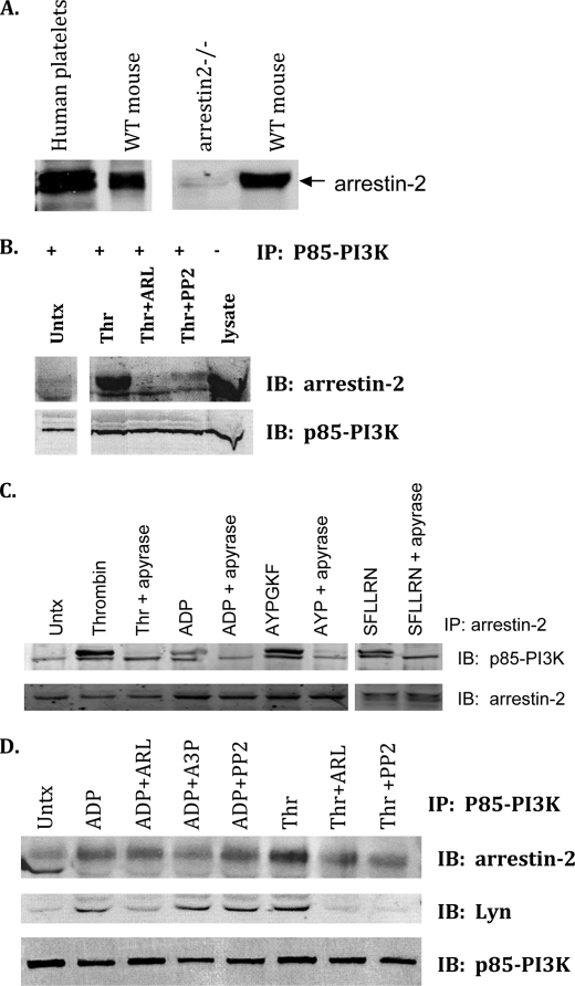
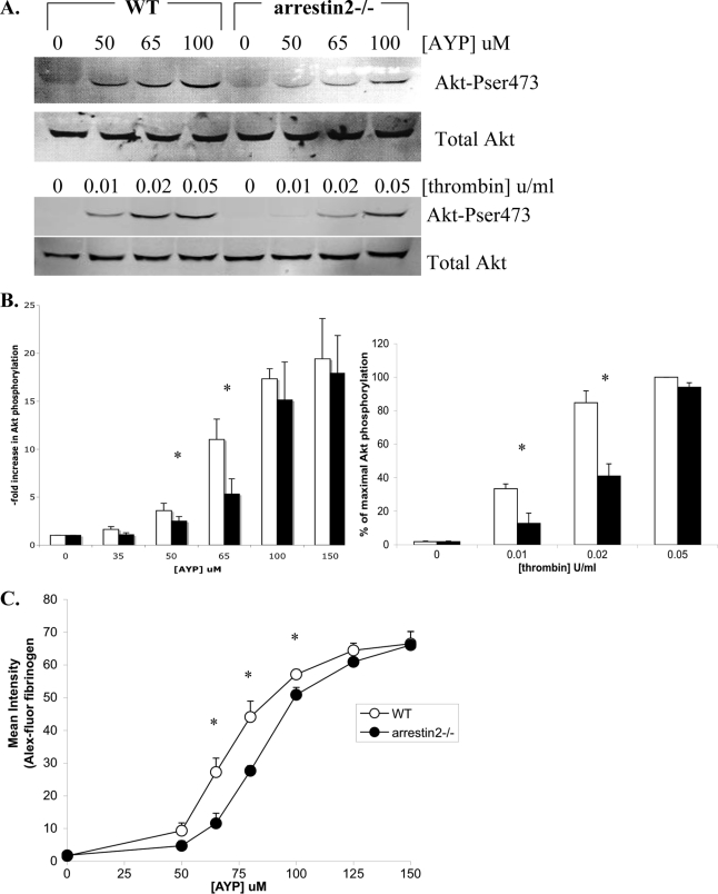
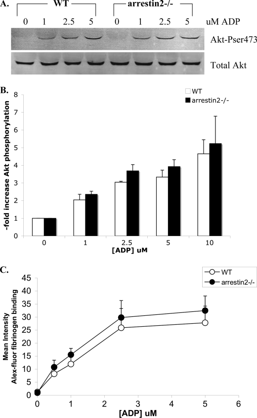
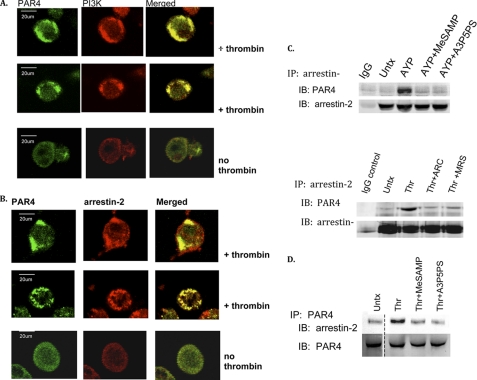
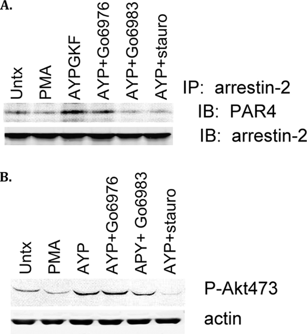
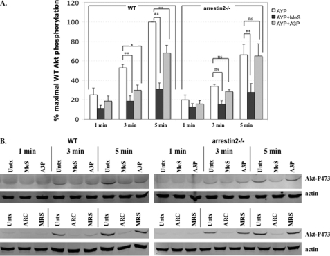
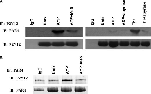
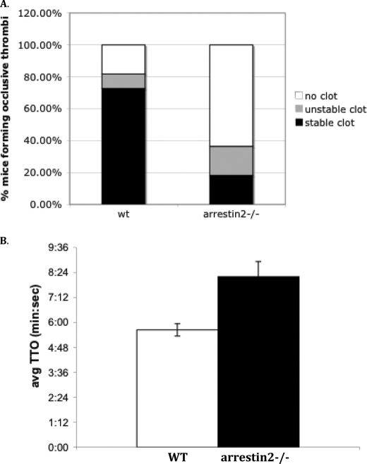
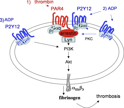
Arrestins can facilitate desensitization or signaling by G protein-coupled receptors (GPCR) in many cells, but their roles in platelets remain uncharacterized. Because of recent reports that arrestins can serve as scaffolds to recruit phosphatidylinositol-3 kinases (PI3K)s to GPCRs, we sought to determine whether arrestins regulate PI3K-dependent Akt signaling in platelets, with consequences for thrombosis. Co-immunoprecipitation experiments demonstrate that arrestin-2 associates with p85 PI3Kα/β subunits in thrombin-stimulated platelets, but not resting cells. The association is inhibited by inhibitors of P2Y12 and Src family kinases (SFKs). The function of arrestin-2 in platelets is agonist-specific, as PAR4-dependent Akt phosphorylation and fibrinogen binding were reduced in arrestin-2 knock-out platelets compared with WT controls, but ADP-stimulated signaling to Akt and fibrinogen binding were unaffected. ADP receptors regulate arrestin recruitment to PAR4, because co-immunoprecipitates of arrestin-2 with PAR4 are disrupted by inhibitors of P2Y1 or P2Y12. P2Y1 may regulate arrestin-2 recruitment to PAR4 through protein kinase C (PKC) activation, whereas P2Y12 directly interacts with PAR4 and therefore, may help to recruit arrestin-2 to PAR4. Finally, arrestin2(-/-) mice are less sensitive to ferric chloride-induced thrombosis than WT mice, suggesting that arrestin-2 can regulate thrombus formation in vivo. In conclusion, arrestin-2 regulates PAR4-dependent signaling pathways, but not responses to ADP alone, and contributes to thrombus formation in vivo.


Figures

FIGURE 1.
FIGURE 1.
Arrestin-2 expression and complex formation in mouse and human platelets. A, 2 × 10⁷ mouse or human platelets were loaded per lane and immunoblotted with antibody to arrestin-2. B, human platelets were left untreated or stimulated by thrombin (0.1U/ml) for 10 min with or without ARL66096(300 nm) or PP2(50uM), lysed, immunoprecipitated with antibodies to p85-PI3K(Upstate, Temecula CA; 2 μg/ml) and immunoblotted with antibodies to arrestin-2 (Santa Cruz Biotechnology, 1:000). C, platelets were stimulated for 10 min with ADP(10 μm), thrombin(0.1 units/ml), peptides AYPGKF(150uM), or SFLLRN(5 μm), with or without apyrase (1 unit/ml); then lysed and immunoprecipitated with antibody to arrestin-2 and immunoblotted with anti-p85-PI3K. D, human platelets treated with ADP or thrombin as in C, with or without ARL66096 (300 nm), A3P5PS(300 μm), or PP2(50um) were immunoprecipitated with antibody to p85-PI3K (2 μg/ml) and immunoblotted with antibodies to arrestin-2, Lyn kinase, or p85-PI3K. Each of the figures shown is representative of results from a minimum of three separate experiments.
FIGURE 2.
FIGURE 2.
Akt phosphorylation and fibrinogen binding in response to PAR4 agonist or thrombin in WT and arrestin2−/− platelets. A, platelets (2 × 10⁷/lane) from WT or arrestin-2−/− mice were stimulated for 5 min at 37 °C with the indicated concentration of AYPGKF, lysed, resolved by SDS-PAGE, and immunoblotted with phosphospecific antibody to p-Akt473 or total Akt. B, average ± S.E. of three or more experiments at each concentration as in A, quantified by densitometry, is shown. White bars are WT, black are arrestin-2−/−. * indicates a significant difference between arrestin-2−/− and WT platelets is detected by 2-tailed, paired Student's t test, with p ≤ 0.05. C, platelets from WT or arrestin-2−/− mice (4 × 10⁷/ml) were stimulated with the indicated concentration of AYPGKF together with AlexaFluor488-conjugated fibrinogen, then fixed and analyzed by flow cytometry. Shown is the mean fluorescence intensity, averaged over three experiments ± S.E. * indicates significant difference between arrestin-2−/− and WT platelets is detected by 2-tailed, paired Student's t test, with p ≤ 0.05.
FIGURE 3.
FIGURE 3.
Akt phosphorylation and fibrinogen binding in response to ADP in WT and arrestin-2−/− platelets. A, platelets from WT or arrestin-2−/− mice were stimulated with the indicated concentration of ADP and immunoblotted for p-Akt473 or total Akt as in Fig. 2. B, average ± S.E. of three experiments as in A, quantified by densitometry is shown. C, platelets from WT or arrestin-2−/− mice were stimulated with the indicated concentration of ADP and analyzed for fibrinogen binding by flow cytometry as in Fig. 2. Shown is the mean fluorescence intensity, averaged over three experiments ± S.E.
FIGURE 4.
FIGURE 4.
Immunofluorescence localization of PAR4, arrestin-2, and p85-PI3K (A and B) and co-immunoprecipitation of PAR4 and arrestin-2 (C and D). A, mouse megakaryocytes were differentiated in culture, grown on Fluorodishes, and incubated in the presence (upper panels) versus absence (lower panels) of thrombin (2 units/ml) for 10 min at 37 °C. Cells were then incubated with FITC-conjugated antibody to PAR4 and rhodamine-conjugated antibody to p85-PI3K (A) or rhodamin-conjugated antibody to arrestin-2 (B), fixed, and slides evaluated at 40× magnification on an Olympus Confocal microscope. 2-Color merge is shown in yellow. C, human platelets (4 × 10⁸/lane) were treated with AYPGKF (150uM) for 5 min at 37 °C with or without 2MeSAMP (100 μm), A3P5PS (300 μm), ARC69931MX (300 nm), or MRS2179 (100 μm), then immunoprecipitated with either IgG control or antibody to arrestin-2. Precipitates were immunoblotted with anti-PAR4 or -arrestin-2 antibodies. D, human platelets were incubated with/without thrombin (0.1 unit/ml) for 5 min at 37 °C, then immunoprecipitated with antibody to PAR4 and immunoblotted with antibodies to arrestin-2 or PAR4.
FIGURE 5.
FIGURE 5.
Effect of PKC inhibition on arrestin-2 association with PAR4 and Akt phosphorylation. A, human platelets (4 × 10⁸/lane) were preincubated with or without PKC inhibitors Go6983 (1 μm), Go6976(1 μm), or staurosporine (1 μm), then stimulated by PMA (1 μm) or AYPGKF (120 μm) and lysed. Arrestin-2 was immunoprecipitated as in Fig. 4B, then precipitates were immunoblotted for PAR4 or arrestin-2. B, human platelets (2 × 10⁷/lane) treated with inhibitors as in A were lysed and immunoblotted with phosphospecific antibody to Akt-Ser-473 or actin. Results shown are representative of two experiments with similar results.
FIGURE 6.
FIGURE 6.
Effect of P2Y1 and P2Y12 inhibition on Akt phosphorylation in WT versus arrestin-2−/− platelets. Platelets from WT or arrestin-2−/− mice (2 × 10⁷/lane) were incubated for the indicated amount of time with 75 μm AYPGKF without inhibitor (white bars), with 2MeSAMP (100 μm) (black bars) or with A3P5PS(300 μm) (gray bars), then lysed and immunoblotted for Akt phosphorylation of Ser-473 as in Fig. 2A. Immunoblots were scanned by densitometry and expressed as % maximal Akt phosphorylation detected in WT platelets stimulated for 5 min (A). The average of four experiments ± S.E. is shown. A significant difference between drug-treated and untreated sample at that time point is denoted by * with p ≤ 0.05 or ** with p ≤ 0.01 using 2-way ANOVA with Bonferroni post-test analysis. Representative immunoblots probed with phospho-Akt antibody (upper blots) and re-probed with antibody to actin (lower blots) are shown in B. Also shown are representative immunoblots of AYPGKF-stimulated platelets in the presence or absence of ARC69931MX (300 nm), or MRS2179 (100 μm) to inhibit P2Y12 and P2Y1, respectively. The same results were obtained in two additional experiments using these inhibitors.
FIGURE 7.
FIGURE 7.
Agonist-dependent association of PAR4 and P2Y12. A, human platelets (4 × 10⁸/lane) were treated with AYPGKF (150uM), ADP (10 μm), or Thrombin (0.1 unit) for 5 min at 37 °C with/without 2MeSAMP (100 μm) or Apyrase (1 unit/ml), then immunoprecipitated with either IgG control or antibody to P2Y12 (2 μg/ml). Precipitates were immunoblotted with anti-PAR4 antibodies (1:1000). B, human platelets treated as above were immunoprecipitated with antibody to PAR4 (2 μg/ml), then immunoblotted with antibody to P2Y12 (1:1000).
FIGURE 8.
FIGURE 8.
Arterial thrombus formation in WT and arrestin-2−/− mice. A, 10% ferric chloride-soaked filter paper was applied for 2 min and 15 s (2′15”) to carotid arteries of pentobarbitol-sedated wild type or arrestin-2−/− mice and the carotid arterial flow rate was measured using a Doppler flow probe. The percentage of each genotype forming stable thrombi that completely impeded flow rate for 30 min is shown in black, the percentage forming unstable thrombi are shown in gray, and the percentage with no occlusion is shown in white. The number of stable occlusions formed differs between WT and arrestin-2−/− mice, with p = 0.03 (two-tailed Fisher's exact probability test). The results of 11 WT and 11 arrestin-2−/− mice are shown. B, 5% ferric chloride-soaked filter paper was applied for 3 min to carotid arteries of sedated mice and flow rate was recorded as described: time to occlusive thrombus formation was recorded for 10 mice of each genotype. The mean time to occlusions differ between the two genotypes of mice with p = 0.005 (unpaired 2-tailed Student's t test).
FIGURE 9.
FIGURE 9.
Thrombin-dependent signaling in platelets (1) requires coincident signaling by ADP(2) for maximal Akt phosphorylation and fibrinogen binding. Part of the requirement for coincident activation of ADP receptors is due to P2Y1-mediated recruitment of arrestin-2 to the PAR4 receptor complex. P2Y12 activation is also required for arrestin recruitment to the PAR4 complex. However, Akt phosphorylation mediated by ADP stimulation of P2Y12 alone (in the absence of thrombin-dependent PAR4 activation) is not dependent on arrestin-2 (3).

Similar articles

Cited by

References

    1. Reiter E., Lefkowitz R. J. (2006) Trends Endocrinol. Metab. 17, 159–165 - PubMed
    1. DeWire S. M., Ahn S., Lefkowitz R. J., Shenoy S. K. (2007) Annu. Rev. Physiol. 69, 483–510 - PubMed
    1. Luttrell L. M., Ferguson S. S., Daaka Y., Miller W. E., Maudsley S., Della Rocca G. J., Lin F., Kawakatsu H., Owada K., Luttrell D. K., Caron M. G., Lefkowitz R. J. (1999) Science 283, 655–661 - PubMed
    1. Yang M., He R. L., Benovic J. L., Ye R. D. (2009) Biochem. J. 417, 287–296 - PMC - PubMed
    1. Lodeiro M., Theodoropoulou M., Pardo M., Casanueva F. F., Camiña J. P. (2009) PLoS One 4, e4686. - PMC - PubMed

Publication types

Substances

LinkOut - more resources