

Regulation of heme oxygenase-1 protein expression by miR-377 in combination with miR-217
- PMID: 21106538
- PMCID: PMC3030323
- DOI: 10.1074/jbc.M110.148726
Regulation of heme oxygenase-1 protein expression by miR-377 in combination with miR-217
Abstract
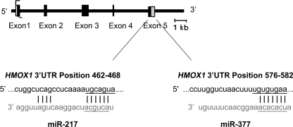
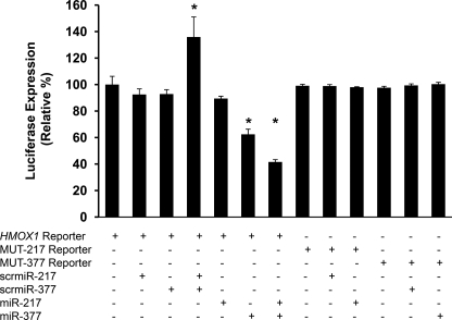
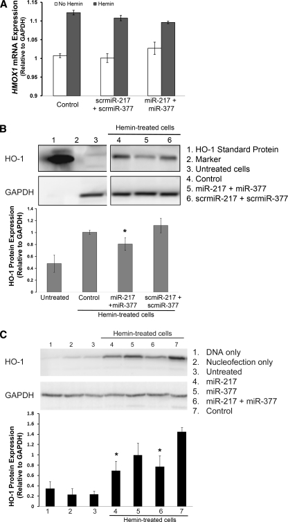
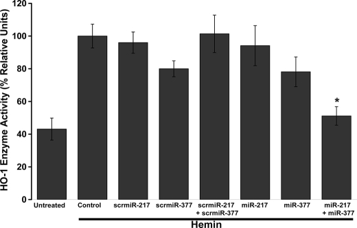
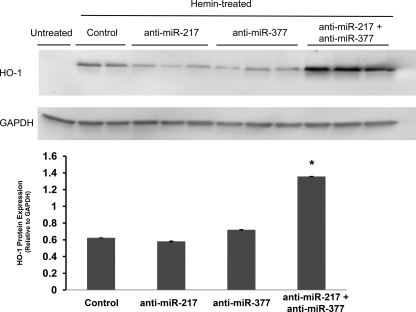
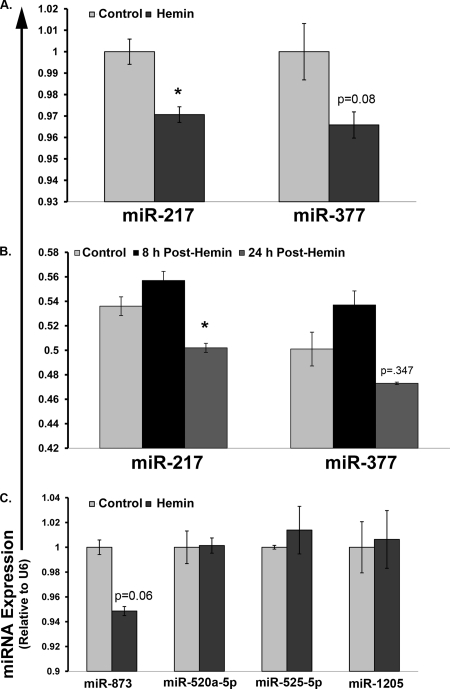
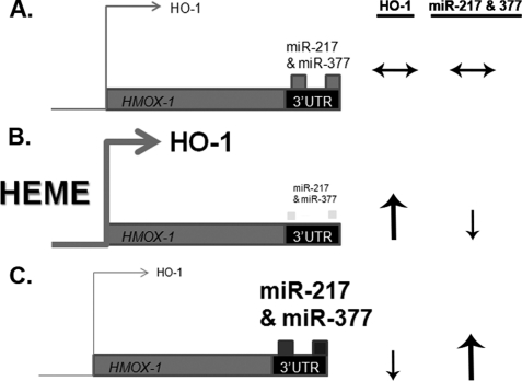
Heme oxygenase-1 (HO-1) enzyme plays a critical role in metabolizing the excess heme generated during hemolysis. Our previous studies suggested that during intravascular hemolysis the expression of HO-1 protein is not sufficient to reduce the oxidative burden of free heme in the vasculature. This led us to hypothesize that a post-translational mechanism of control exists for HO-1 expression. Micro-RNAs (miRNA) affect gene expression by post-transcriptional gene regulation of transcripts. We performed in silico analysis for the human HMOX1-3' untranslated region (3' UTR) and identified candidate miRNA binding sites. Two candidate miRNAs, miR-377 and miR-217, were cloned and co-transfected with a luciferase vector containing the human HMOX1-3'UTR region. The combination of miR-377 and miR-217 produced a 58% reduction in HMOX1-3'UTR luciferase reporter expression compared with controls. The same constructs were then used to assess how overexpression of miR-217 and miR-377 affected HO-1 levels after induction with hemin. Cells transfected with the combination of miR-377 and miR-217 exhibited no change in HMOX1 mRNA levels, but a significant reduction in HMOX1 (HO-1) protein expression and enzyme activity compared with non-transfected hemin-stimulated controls. Transfection with either miR-377 or miR-217 alone did not produce a significant decrease in HO-1 protein expression or enzyme activity. Knockdown of miR-217 and miR-377 in combination leads to up-regulation of HO-1 protein. Exposure to hemin induced a significant reduction in miR-217 expression and a trend toward decreased miR-377 expression in two different cells lines. In summary, these data suggests that the combination of miR-377 and miR-217 help regulate HO-1 protein expression in the presence of hemin.
Figures
Similar articles
-
MicroRNA-196 represses Bach1 protein and hepatitis C virus gene expression in human hepatoma cells expressing hepatitis C viral proteins.Hepatology. 2010 May;51(5):1494-504. doi: 10.1002/hep.23401. Hepatology. 2010. PMID: 20127796 Free PMC article.
-
The let-7 microRNA enhances heme oxygenase-1 by suppressing Bach1 and attenuates oxidant injury in human hepatocytes.Biochim Biophys Acta. 2012 Nov-Dec;1819(11-12):1113-22. doi: 10.1016/j.bbagrm.2012.06.001. Epub 2012 Jun 12. Biochim Biophys Acta. 2012. PMID: 22698995 Free PMC article.
-
Hemin inhibits NO production by IL-1β-stimulated human astrocytes through induction of heme oxygenase-1 and reduction of p38 MAPK activation.J Neuroinflammation. 2010 Sep 7;7:51. doi: 10.1186/1742-2094-7-51. J Neuroinflammation. 2010. PMID: 20822529 Free PMC article.
-
Heme oxygenase-1 modulates microRNA expression in cultured astroglia: implications for chronic brain disorders.Glia. 2015 Jul;63(7):1270-84. doi: 10.1002/glia.22823. Epub 2015 Mar 25. Glia. 2015. PMID: 25820186
-
Dysregulation of a Heme Oxygenase-Synuclein Axis in Parkinson Disease.NeuroSci. 2022 May 20;3(2):284-299. doi: 10.3390/neurosci3020020. eCollection 2022 Jun. NeuroSci. 2022. PMID: 39483365 Free PMC article. Review.
Cited by
-
Heme Oxygenase-1 and Prostate Cancer: Function, Regulation, and Implication in Cancer Therapy.Int J Mol Sci. 2024 Aug 24;25(17):9195. doi: 10.3390/ijms25179195. Int J Mol Sci. 2024. PMID: 39273143 Free PMC article. Review.
-
MicroRNA-217 promotes ethanol-induced fat accumulation in hepatocytes by down-regulating SIRT1.J Biol Chem. 2012 Mar 23;287(13):9817-9826. doi: 10.1074/jbc.M111.333534. Epub 2012 Feb 3. J Biol Chem. 2012. PMID: 22308024 Free PMC article.
-
Translocation of heme oxygenase-1 to mitochondria is a novel cytoprotective mechanism against non-steroidal anti-inflammatory drug-induced mitochondrial oxidative stress, apoptosis, and gastric mucosal injury.J Biol Chem. 2011 Nov 11;286(45):39387-402. doi: 10.1074/jbc.M111.279893. Epub 2011 Sep 9. J Biol Chem. 2011. PMID: 21908612 Free PMC article.
-
Hemin controls T cell polarization in sickle cell alloimmunization.J Immunol. 2014 Jul 1;193(1):102-10. doi: 10.4049/jimmunol.1400105. Epub 2014 May 30. J Immunol. 2014. PMID: 24879794 Free PMC article. Clinical Trial.
-
Heme oxygenase, a novel target for the treatment of hypertension and obesity?Am J Physiol Regul Integr Comp Physiol. 2012 Jan 15;302(2):R207-14. doi: 10.1152/ajpregu.00517.2011. Epub 2011 Nov 9. Am J Physiol Regul Integr Comp Physiol. 2012. PMID: 22071158 Free PMC article. Review.
References
-
- Kumar S., Bandyopadhyay U. (2005) Toxicol. Lett. 157, 175–188 - PubMed
-
- Sugishima M., Omata Y., Kakuta Y., Sakamoto H., Noguchi M., Fukuyama K. (2000) FEBS Lett. 471, 61–66 - PubMed
-
- Otterbein L. E., Soares M. P., Yamashita K., Bach F. H. (2003) Trends Immunol. 24, 449–455 - PubMed
-
- Wagener F. A., Volk H. D., Willis D., Abraham N. G., Soares M. P., Adema G. J., Figdor C. G. (2003) Pharmacol. Rev. 55, 551–571 - PubMed
Publication types
MeSH terms
Substances
Grants and funding
LinkOut - more resources
Full Text Sources
Other Literature Sources
