. 2011 Feb 4;286(5):3405-17.
doi: 10.1074/jbc.M110.190637. Epub 2010 Nov 24.

Heterocomplexes of mannose-binding lectin and the pentraxins PTX3 or serum amyloid P component trigger cross-activation of the complement system

Affiliations

Heterocomplexes of mannose-binding lectin and the pentraxins PTX3 or serum amyloid P component trigger cross-activation of the complement system

Ying Jie Ma et al. J Biol Chem. .

Abstract

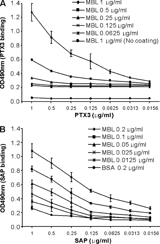
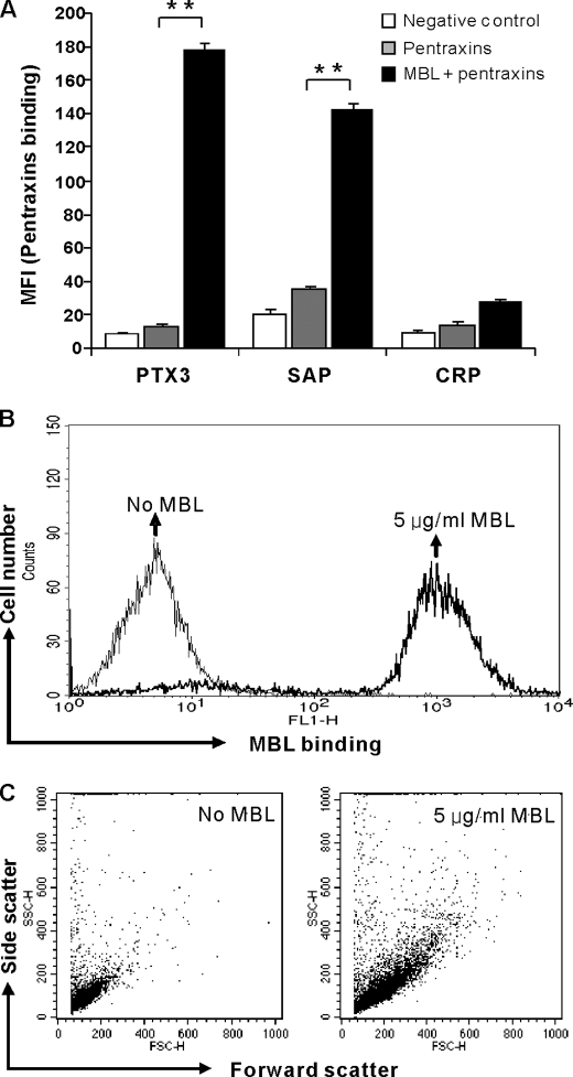
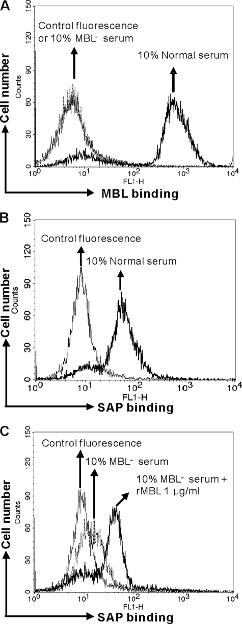
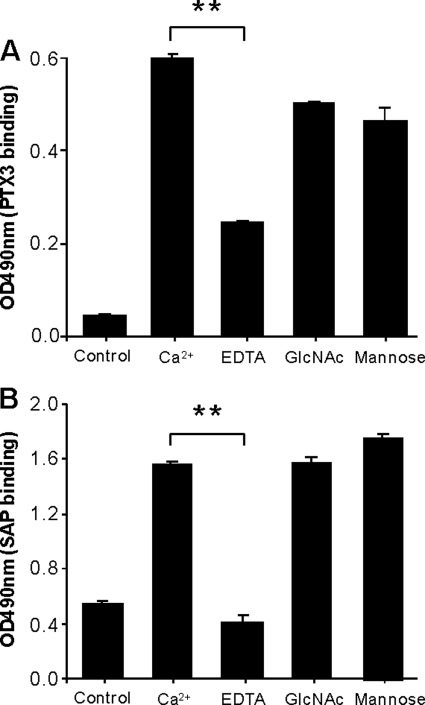
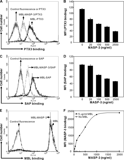
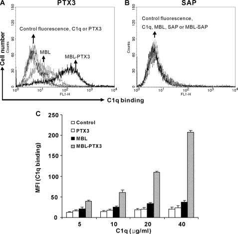
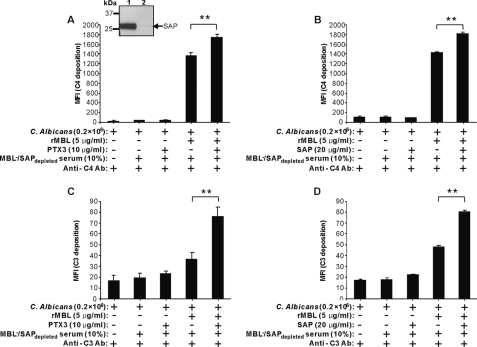
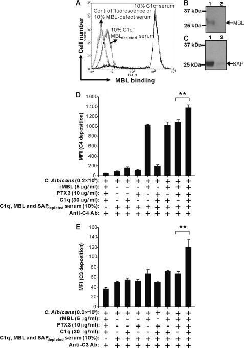
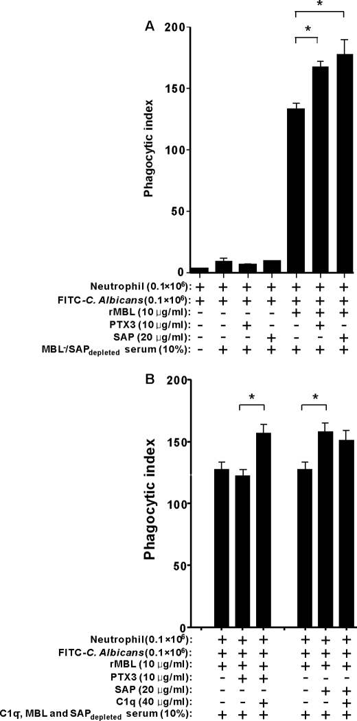
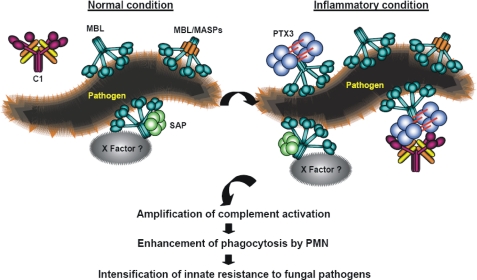
The long pentraxin 3 (PTX3), serum amyloid P component (SAP), and C-reactive protein belong to the pentraxin family of pattern recognition molecules involved in tissue homeostasis and innate immunity. They interact with C1q from the classical complement pathway. Whether this also occurs via the analogous mannose-binding lectin (MBL) from the lectin complement pathway is unknown. Thus, we investigated the possible interaction between MBL and the pentraxins. We report that MBL bound PTX3 and SAP partly via its collagen-like domain but not C-reactive protein. MBL-PTX3 complex formation resulted in recruitment of C1q, but this was not seen for the MBL-SAP complex. However, both MBL-PTX3 and MBL-SAP complexes enhanced C4 and C3 deposition and opsonophagocytosis of Candida albicans by polymorphonuclear leukocytes. Interaction between MBL and PTX3 led to communication between the lectin and classical complement pathways via recruitment of C1q, whereas SAP-enhanced complement activation occurs via a hitherto unknown mechanism. Taken together, MBL-pentraxin heterocomplexes trigger cross-activation of the complement system.


Figures

FIGURE 1.
FIGURE 1.
Dose-dependent binding of PTX3 and SAP to MBL in solid phase ELISA. A, binding of PTX3 to MBL is shown. Microtiter plates coated with or without mannan (10 μg/ml) were incubated with the indicated concentrations of MBL before the addition of PTX3 in 2-fold serial dilutions. B, binding of SAP to MBL is shown. To reduce nonspecific background, MBL or BSA (control) was coated directly to microtiter plates in various concentrations and incubated with SAP in 2-fold serial dilutions. Binding of the pentraxins to the microtiter plates was assessed by anti-PTX3 and anti-SAP antibodies. Results are represented as the mean ± S.E. from three independent experiments with duplicates.
FIGURE 2.
FIGURE 2.
Binding of pentraxins to C. albicans in the presence of MBL. A, C. albicans was incubated with PTX3 (10 μg/ml), SAP (20 μg/ml), or CRP (20 μg/ml) in the absence or presence of MBL (5 μg/ml) and detected by FACS analysis. As controls, MBL binding (B) and MBL-dependent agglutination (C) were assessed. The MFI was used to assess PTX3, SAP, CRP, and MBL binding. Agglutination was assessed by the change in forward and scatter morphology. Data are expressed as the mean ± S.E. from triplicate experiments. Results are representative of at least six independent experiments. The asterisks indicate the statistical significance versus controls: **, p < 0.01.
FIGURE 3.
FIGURE 3.
Formation and presence of MBL-SAP complexes in normal serum. A, detection of serum MBL on C. albicans is shown. B, detection of serum SAP binding to C. albicans is shown. C, detection of serum SAP binding in MBL-defect (MBL) serum with or without exogenous MBL is shown. The MFI was used to assess SAP or MBL binding. Results are representative of three independent experiments.
FIGURE 4.
FIGURE 4.
Effect of EDTA, GlcNAc, and mannose on the binding of PTX3 or SAP to MBL. A, microtiter plates coated with or without (control) anti-MBL monoclonal Ab (HYB 131-01) were incubated with MBL (1 μg/ml) before the addition of PTX3 (1 μg/ml) alone in TBS-T buffer containing Ca2+ (2.5 mm) or the buffer containing EDTA (10 mm), GlcNAc (0.1 m), or mannose (0.1 m), respectively. Bound PTX3 was detected as depicted above. B, microtiter plates coated with MBL (0.2 μg/ml) or control BSA (0.2 μg/ml) followed by incubation of SAP (1 μg/ml) in TBS-T buffer containing Ca2+ (2.5 mm) or the buffer containing EDTA (10 mm), GlcNAc (0.1 m), and mannose (0.1 m), respectively are shown. Data are expressed as the mean ± S.E. from triplicate experiments. Results are representative of three independent experiments. The asterisks indicate the statistical significance versus controls: **, p < 0.01.
FIGURE 5.
FIGURE 5.
Involvement of collagen-like domain of MBL in MBL-PTX3 or MBL-SAP complex formation. C. albicans was incubated with or without MBL (5 μg/ml) followed by incubation with PTX3 (10 μg/ml) (A) or SAP (20 μg/ml) (C) in the absence or presence of MASP-3 (5000 ng/ml). Dose-dependent inhibitory effect of MASP-3 was determined by incubating PTX3 (10 μg/ml) (B) or SAP (20 μg/ml) (D) in the presence of MBL (5 μg/ml) with increasing concentrations of MASP-3. E, control for MBL binding was determined using MBL (5 μg/ml) in the absence or presence of MASP-3 (5000 ng/ml). F, control for MASP-3 binding was determined with or without MBL in increasing concentrations of MASP-3. The MFI was used to assess PTX3, SAP, MBL, or MASP-3 binding. Results (A, C, E, and F) are representative of three independent experiments. B and D are expressed as the mean ± S.E. from triplicate experiments and are representative of two independent experiments that yield similar results.
FIGURE 6.
FIGURE 6.
C1q binding to MBL-PTX3 complexes but not MBL-SAP complexes. C. albicans treated with MBL (5 μg/ml), PTX3 (10 μg/ml) (A), SAP (20 μg/ml) (B), or mixtures of both were incubated with C1q (30 μg/ml). Bound C1q was detected. As a control, C. albicans was incubated with C1q alone. C, in some experiments, increasing concentrations of C1q were applied to determine dose dependence. The MFI was used to assess C1q binding. Results A and B are representative of three independent experiments. Data C are expressed as the mean ± S.E. from triplicate experiments. Results are representative of two independent experiments that yield similar results.
FIGURE 7.
FIGURE 7.
Enhancement of C4 and C3 deposition by MBL-PTX3 or SAP complex formation on C. albicans. MBL-PTX3 or MBL-SAP complexes were first established on C. albicans as described above before the addition of 10% MBL/SAPdepleted serum. C4 and C3 deposition were assessed. As controls, the addition of MBL and PTX3 alone was applied. The inset shows depletion of SAP from MBL serum using DNA-cellulose. Lane 1, before depletion; lane 2, after depletion. The MFI was used to assess C4 and C3 deposition. Results are presented as the mean ± S.E. of samples analyzed in triplicate. Results are representative of three independent experiments. The asterisks indicate the statistical significance versus controls: **, p < 0.01.
FIGURE 8.
FIGURE 8.
C1q-dependent enhancement of C4 and C3 deposition by MBL-PTX3 complexes on C. albicans. MBL was depleted from C1q serum and assessed by FACS (A) and Western blot (B), then SAP was depleted and assessed by Western blot (C). Lane 1, before depletion; lane 2, after depletion. The MBL-PTX3 complex was first established on C. albicans as described above followed by induction of C4 and C3 deposition in 10% C1q/MBL and SAPdepleted serum with or without exogenous C1q. D, and E, C4 and C3 deposition were assessed by FACS. As controls, the addition of MBL, PTX3 alone, or mixtures of both were applied. The MFI was used to assess C4 and C3 deposition. Results are presented as the mean ± S.E. of samples analyzed in triplicate. Results are representative of three independent experiments. The asterisks indicate the statistical significance versus controls: **, p < 0.01.
FIGURE 9.
FIGURE 9.
C1q-dependent enhancement of phagocytosis by human neutrophils. A, complement dependent-phagocytosis of C. albicans by human neutrophils is shown. C. albicans were labeled with FITC and preincubated with or without MBL, PTX3, or SAP or a combination of the molecules to induce complement activation in MBL/SAPdepleted serum. B, effect of C1q on phagocytosis of C. albicans induced by MBL-PTX3 complex is shown. C1q/MBL and SAPdepleted serum was used as a complement source to opsonize FITC-labeled C. albicans after incubation with or without MBL, PTX3, SAP, C1q, or a combination of the molecules. Phagocytosis was analyzed by FACS. The bars represent values expressed as the average opsonophagocytosis ± S.E. of six donors (A) or three donors (B) measured on three different days. The asterisk indicates the statistical significance versus controls: *, p < 0.05.
FIGURE 10.
FIGURE 10.
A proposed model of the novel complement amplification mechanisms created by MBL-PTX3 and MBL-SAP complexes. MBL-PTX3 interaction induces amplification of complement activation via C1q recruitment and the classical pathway, resulting in increased deposition of complement factors. MBL-SAP interaction induces amplification of complement activation via a hitherto unidentified X serum factor(s) without involvement of C1q.

Similar articles

Cited by

References

    1. Takahashi K., Ip W. E., Michelow I. C., Ezekowitz R. A. (2006) Curr. Opin. Immunol. 18, 16–23 - PMC - PubMed
    1. Garred P., Larsen F., Seyfarth J., Fujita R., Madsen H. O. (2006) Genes Immun 7, 85–94 - PubMed
    1. Thiel S., Vorup-Jensen T., Stover C. M., Schwaeble W., Laursen S. B., Poulsen K., Willis A. C., Eggleton P., Hansen S., Holmskov U., Reid K. B., Jensenius J. C. (1997) Nature 386, 506–510 - PubMed
    1. Chen C. B., Wallis R. (2004) J. Biol. Chem. 279, 26058–26065 - PubMed
    1. Takahashi M., Iwaki D., Kanno K., Ishida Y., Xiong J., Matsushita M., Endo Y., Miura S., Ishii N., Sugamura K., Fujita T. (2008) J. Immunol. 180, 6132–6138 - PubMed

Publication types

MeSH terms

LinkOut - more resources