Review
. 2011 Mar 1;3(3):a000638.
doi: 10.1101/cshperspect.a000638.

The nucleolus

Affiliations
Review

The nucleolus

Thoru Pederson. Cold Spring Harb Perspect Biol. .

Abstract

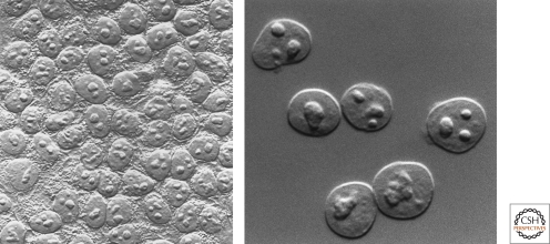
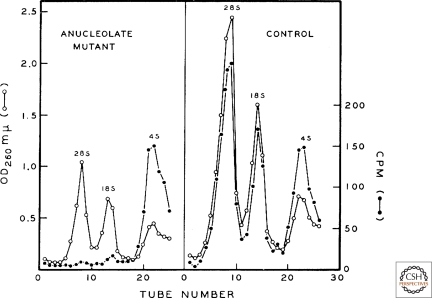
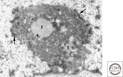
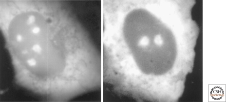
When cells are observed by phase contrast microscopy, nucleoli are among the most conspicuous structures. The nucleolus was formally described between 1835 and 1839, but it was another century before it was discovered to be associated with a specific chromosomal locus, thus defining it as a cytogenetic entity. Nucleoli were first isolated in the 1950s, from starfish oocytes. Then, in the early 1960s, a boomlet of studies led to one of the epochal discoveries in the modern era of genetics and cell biology: that the nucleolus is the site of ribosomal RNA synthesis and nascent ribosome assembly. This epistemologically repositioned the nucleolus as not merely an aspect of nuclear anatomy but rather as a cytological manifestation of gene action-a major heuristic advance. Indeed, the finding that the nucleolus is the seat of ribosome production constitutes one of the most vivid confluences of form and function in the history of cell biology. This account presents the nucleolus in both historical and contemporary perspectives. The modern era has brought the unanticipated discovery that the nucleolus is plurifunctional, constituting a paradigm shift.


Figures

Figure 1.
Figure 1.
Looking at the nucleolus. Nucleoli observed in HeLa cells (left) and isolated HeLa cell nuclei (right) by differential interference microscopy. (Images courtesy of David L. Spector [left] and Thoru Pederson [right].)
Figure 2.
Figure 2.
A portal to the modern era: the dependence of ribosomal RNA synthesis on the nucleolus. Anucleolate (left) or wild-type (right) Xenopus laevis embryos at the neurula stage were incubated with C14-labeled carbon dioxide and RNA was extracted 20 hours later, when both groups of embryos were still morphologically and physiologically indistinguishable. Shown are sucrose gradient sedimentation profiles of 28S and 18S rRNA and transfer RNA. This finding was a keystone in establishing the role of the nucleolus in the biosynthesis of ribosomes. (Reproduced from Brown DC and Gurdon JB 1964. Proc Natl Acad Sci USA 51: 139–146, by kind permission of Donald D. Brown, Carnegie Institution for Science, Washington, D.C.)
Figure 3.
Figure 3.
The tripartite organization of the nucleolus. Electron micrograph of a mouse fibroblast nucleolus. (f) fibrillar center, (d) dense fibrillar component, (g) granular component, (arrows) perinucleolar heterochromatin, (*) denotes the presence of dense fibrillar component material within the fibrillar center, which is occasionally observed. (Reprinted from Trends in Cell Biol 13: 517–525, Raška, I. © 2003, with permission from Elsevier.)
Figure 4.
Figure 4.
The nucleolus and signal recognition particle biosynthesis. Nucleolar localization of GFP-tagged SRP19 (left) and SRP68 (right) proteins in rat NRK cells. Cytoplasmic localization is also observed, as expected from the known steady-state localization of the signal recognition particle. Similar results were obtained with GFP-tagged SRP72 protein, whereas the SRP54 protein assembles with the nascent signal recognition particle in the cytoplasm (Politz et al. 2000; Sommerville et al. 2005). Reproduced from Politz et al. 2000, with permission from the Proceedings of the National Academy of Sciences.
Figure 5.
Figure 5.
The nucleolar granular component contains both RNA-rich and RNA-deficient particles. Electron spectroscopic imaging (ESI) is a mode of energy loss measurement in electron microscopy that can be tuned to record beam collisions with orbital electrons of nitrogen or phosphorus in the specimen. Using the empirical chemical formulas of protein and nucleic acid with respect to these two elements, ESI allows one to construct a map of protein-rich versus nucleic-acid-rich objects. Shown is an ESI image from a portion of a human neuroblastoma cell nucleus spanning a region of the nucleoplasm and part of a nucleolus. The protein-rich and nucleic-acid-rich regions have been colored blue and yellow, respectively. (DCh) decondensed chromatin, (CCh) condensed chromatin, (PML) promyelocytic leukemia body, (GC) granular component of the nucleolus. The PML body (entirely blue, i.e., protein) serves as a valuable internal control, as it is known not to contain appreciable nucleic acid. The granular component of the nucleolus, once thought to be a uniform zone of nascent ribosomes, contains an interspersed landscape of nucleic-acid-rich versus nucleic-acid-deficient particles. ESI cannot distinguish between DNA and RNA, but since several independent methods have detected no DNA in the nucleolar granular component, the nucleic-acid-rich particles are plausibly nascent ribosomes, perhaps with a contribution from assembling signal recognition particles, whereas the nucleic-acid-deficient, protein-rich particles may be heterotypic complexes of cell-cycle regulatory proteins, as discussed. (Reprinted from Politz JC et al. 2005, Mol Biol Cell 16: 3401–3410.)

Similar articles

Cited by

References

    1. Alberts B, Johnson A, Lewis J, Raff M, Roberts K, Walter P 2002. Molecular Biology of the Cell, 4th edition Garland Science, New York: p. 332
    1. Andersen JS, Lyon CE, Fox AH, Leung AK, Lam YW, Steen H, Mann M, Lamond AI 2002. Directed proteomic analysis of the human nucleolus. Curr Biol 12: 1–11 - PubMed
    1. Bachellerie JP, Cavaillé J, Hüttenhofer A 2002. The expanding RNA world. Biochimie 84: 775–790 - PubMed
    1. Boisvert FM, van Koningsbruggen S, Navascués J, Lamond AI 2007. The multifunctional nucleolus. Nat Rev Mol Cell Biol 8: 574–585 - PubMed
    1. Boyne JR, Whitehouse A 2006. Nucleolar trafficking is essential for nuclear export of intronless herpesvirus mRNA. Proc Natl Acad Sci USA 103: 15190–15195 - PMC - PubMed

Publication types

MeSH terms

LinkOut - more resources