Review
. 2012 Feb 1;4(2):a003566.
doi: 10.1101/cshperspect.a003566.

Riboswitches and the RNA world

Affiliations
Review

Riboswitches and the RNA world

Ronald R Breaker. Cold Spring Harb Perspect Biol. .

Abstract

Riboswitches are structured noncoding RNA domains that selectively bind metabolites and control gene expression (Mandal and Breaker 2004a; Coppins et al. 2007; Roth and Breaker 2009). Nearly all examples of the known riboswitches reside in noncoding regions of messenger RNAs where they control transcription or translation. Newfound classes of riboswitches are being reported at a rate of about three per year (Ames and Breaker 2009), and these have been shown to selectively respond to fundamental metabolites including coenzymes, nucleobases or their derivatives, amino acids, and other small molecule ligands. The characteristics of some riboswitches suggest they could be modern descendents of an ancient sensory and regulatory system that likely functioned before the emergence of enzymes and genetic factors made of protein (Nahvi et al. 2002; Vitreschak et al. 2004; Breaker 2006). If true, then some of the riboswitch structures and functions that serve modern cells so well may accurately reflect the capabilities of RNA sensors and switches that existed in the RNA World. This article will address some of the characteristics of modern riboswitches that may be relevant to ancient versions of these metabolite-sensing RNAs.


Figures

Figure 1.
Figure 1.
Classes of validated and candidate riboswitches plotted in descending order relative to their frequency in the genomes of ∼700 bacterial species. Classes with at least some biochemical or genetic validation (filled bars) are named for the ligand that is most tightly bound. Multiple compound names indicate that binding site variants exist with altered ligand specificity. Candidate riboswitches whose ligand-binding functions have not been validated (open bars) are named for genes that are commonly associated with the motif.
Figure 2.
Figure 2.
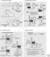
Established or predicted mechanisms of riboswitch-mediated gene regulation. The most common mechanisms are (A) transcription termination, (B) translation initiation, and (C) splicing control (in eukaryotes). More rare mechanisms observed or predicted in some bacterial species include (D) transcription interference or possibly antisense action, (E) dual transcription and translation control, and (F) ligand-dependent self-cleaving ribozyme action. The numbers in a identify steps that are important for kinetically driven riboswitches (Wickiser et al. 2005a,b; Gilbert et al. 2006). Numbers represent (1) folding of the aptamer, (2) docking of the ligand, (3) folding of the expression platform, and (4) speed of RNA polymerase (RNAP). In B, Rho represents the transcriptional terminator protein (Skordalakes and Berger 2003).
Figure 3.
Figure 3.
Kinetic function of an FMN riboswitch. (A) Sequence and secondary structure models for the ribD FMN riboswitch from B. subtilis (Winkler et al. 2002a). The RNA functions as a genetic “OFF” switch wherein FMN binding stabilizes P1 formation, precludes the formation of an antiterminator stem, and permits the formation of a terminator stem that represses gene expression. (B) Simplified kinetic scheme for the function of the ribD FMN riboswitch depicted in A. Steps represented by black and gray arrows lead to termination and full-length mRNA production, respectively. See elsewhere for details (Wickiser et al. 2005b).
Figure 4.
Figure 4.
Riboswitch aptamers that respond to the coenzyme SAM or its metabolic derivative SAH. (A) The chemical structures of SAM and SAH. (B) Three variations of the SAM-I aptamer class. Despite substantial differences in sequence and structural subdomains, SAM-I (Grundy and Henkin 1998), SAM-IV (Weinberg et al. 2008), and SAM-I/SAM-IV (Weinberg et al. 2010) aptamer families carry a common ligand binding core (green-shaded nucleotides), including the identities of nucleotides that directly contact SAM (red bold letters). (C) A second superfamily of SAM-specific riboswitches use either the SAM-II (Corbino et al. 2005) or SAM-V (Poiata et al. 2009) aptamer families, which also carry a common ligand binding core. Annotations are as described for B. (D) Sequence and structural features of SAM-III aptamers (Fuchs et al. 2006), representing the third class of SAM-specific riboswitches. (E) Sequence and structural features of SAM/SAH aptamers (Weinberg et al. 2010), which genetically track like SAM riboswitches but bind SAH more tightly. (F) A distinct class of aptamers that genetically track as SAH riboswitches and selectively bind SAH while strongly discriminating against SAM (Wang et al. 2008).
Figure 5.
Figure 5.
Binding kinetics for simple riboswitches, or complex riboswitches that use cooperative binding or a tandem architecture using riboswitches of from the same class. (A) Dose-response curve for a typical riboswitch carrying a single aptamer that functions perfectly. Note that the plot represents the performance of a population of individual riboswitch molecules where [R] represents the fraction of riboswitches causing gene expression change on ligand binding. Ligand concentration [L] is in arbitrary units, and T50 (Wickiser et al. 2005b) represents the concentration of ligand needed to half-maximally modulate gene expression. (B) Comparison of the dose-response curve for a simple riboswitch (one aptamer and one expression platform) versus a cooperative riboswitch (two aptamers and one expression platform). The curve for the cooperative riboswitch reflects perfect cooperativity between the aptamers and a Hill coefficient (n) of two. Note that [Y], the fraction of riboswitches bound by ligand, is equivalent to [R] if ligand binding always triggers a change in gene expression. (C) Comparison of the dose-response curve for a simple riboswitch versus a tandem arrangement of independently functioning riboswitches of the same class and near identical T50 values. Other annotations are as described in A and B. (Adapted, with permission, from Welz and Breaker 2007.)
Figure 6.
Figure 6.
Architectures and functions of tandem riboswitch arrangements. (A) Cooperative glycine riboswitch system that yields a more digital genetic response in numerous Gram positive bacteria including in the 5′ UTR of the B. subtilis gcvT gene (Mandal et al. 2004). (B) Tandem TPP riboswitches from the 5′ UTR of the thiamin metabolism gene tenA from Bacillus anthracis (Welz and Breaker 2007). (C) Tandem SAM-II and SAM-V riboswitches identified in ocean bacteria such as “Cand. P. ubique” (Poiata et al. 2009). (D) A two-input Boolean NOR logic gate composed of tandem riboswitches for SAM and AdoCbl located in the metE gene from Bacillus clausii (Sudarsan et al. 2006).
Figure 7.
Figure 7.
A possible path for the descent of modern riboswitches from RNA World ribozymes. SAM-dependent RNAs were arbitrarily chosen as an example. (Stage I) Emergence of ribozymes that synthesize SAM. Ribozymes might have coupled methionine to a 5′-terminal ATP moiety or coupled methionine to a free ATP substrate. (Stage II) Utilization of SAM by diverse methyltransferase ribozymes. In a complex RNA World, various methyltransferase ribozymes might have been present that used SAM as a prosthetic group or as a diffusible cofactor. (Stage III) Co-opting ribozyme subdomains to create RNA switches. Allosteric systems that controlled ribozyme function or that controlled the biosynthesis of new RNAs could have made use of variant SAM-binding domains of certain methyltransferase ribozymes. (Stage IV) Perseverance of ancient SAM-binding aptamers and their use in modern riboswitches. (Adapted, with permission, from Breaker 2006.)

References

    1. André G, et al. 2008. S-box and T-box riboswitches and antisense RNA control a sulfur metabolic operon of Clostridium acetobutylicum. Nucleic Acids Res 36: 5955–5969 - PMC - PubMed
    1. Ames TD, Breaker RR 2009. Bacterial riboswitch discovery and analysis. In: The chemical biology of nucleic acids, Meyer G., ed., John Wiley & Sons, Ltd; (in press)
    1. Barrick JE, Breaker RR 2007. The distributions, mechanisms, and structures of metabolite-binding riboswitches. Genome Biol 8: R239. - PMC - PubMed
    1. Benner SA, Ellington AD, Tauer A 1989. Modern metabolism as a palimpsest of the RNA world. Proc Natl Acad Sci USA 86: 7054–7058 - PMC - PubMed
    1. Blount KF, Breaker RR 2006. Riboswitches as antibacterial drug targets. Nat Biotechnol 24: 1558–1564 - PubMed

Publication types

MeSH terms

LinkOut - more resources