

Mitochondrial degradation by autophagy (mitophagy) in GFP-LC3 transgenic hepatocytes during nutrient deprivation
- PMID: 21106691
- PMCID: PMC3043636
- DOI: 10.1152/ajpcell.00056.2010
Mitochondrial degradation by autophagy (mitophagy) in GFP-LC3 transgenic hepatocytes during nutrient deprivation
Abstract
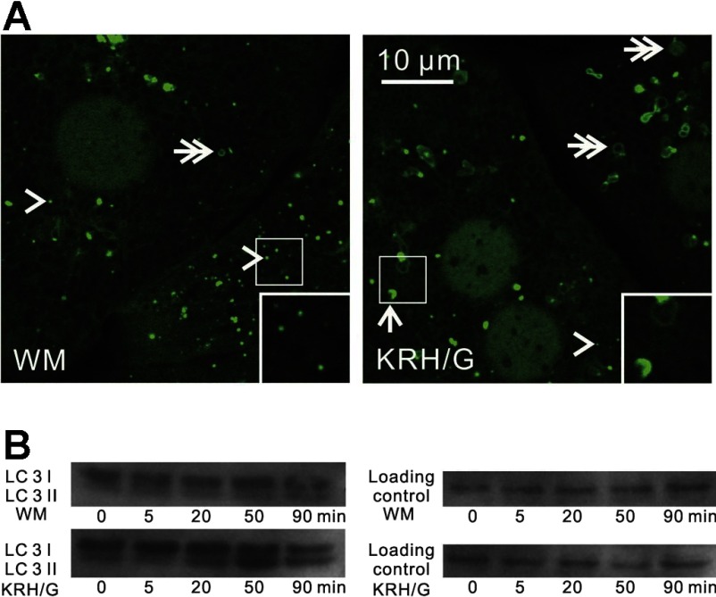
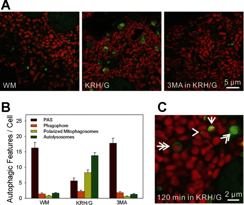
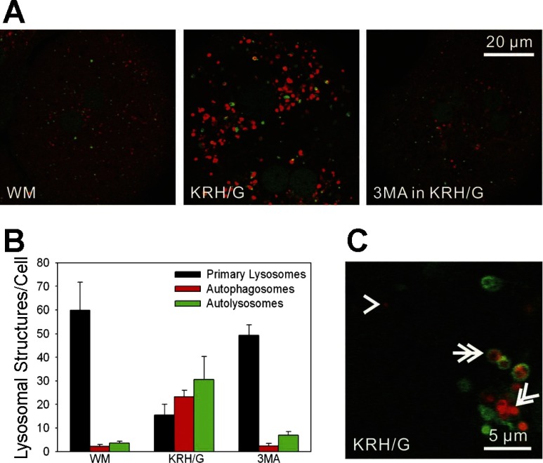
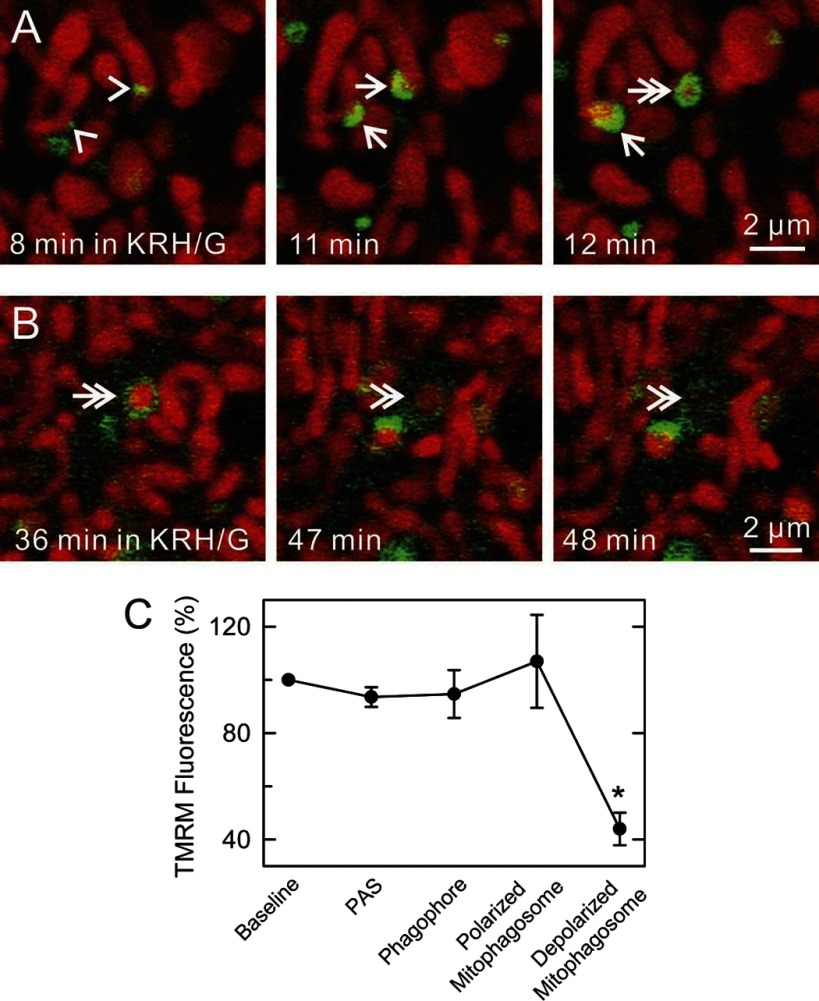
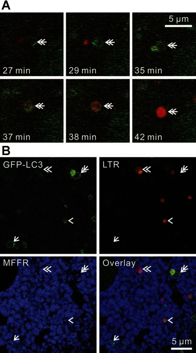
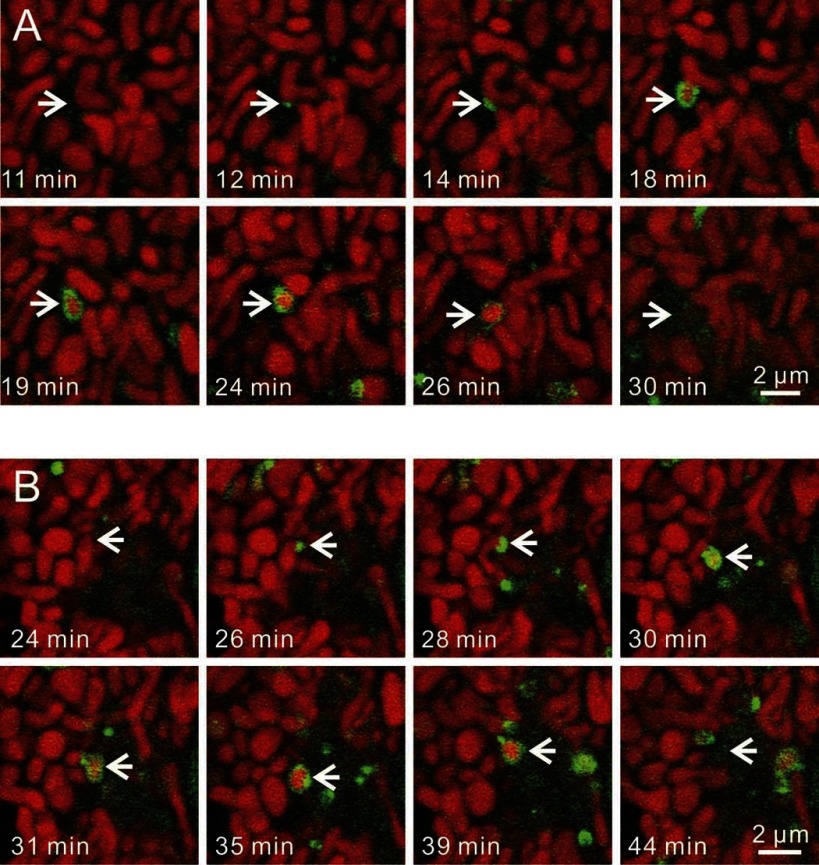
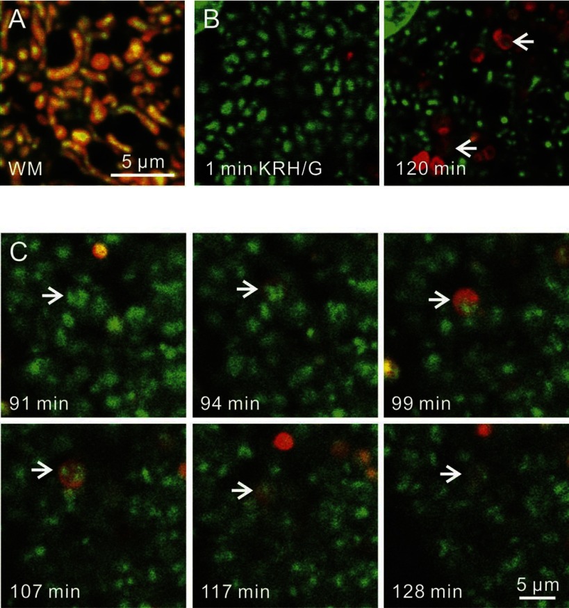
Fasting in vivo and nutrient deprivation in vitro enhance sequestration of mitochondria and other organelles by autophagy for recycling of essential nutrients. Here our goal was to use a transgenic mouse strain expressing green fluorescent protein (GFP) fused to rat microtubule-associated protein-1 light chain 3 (LC3), a marker protein for autophagy, to characterize the dynamics of mitochondrial turnover by autophagy (mitophagy) in hepatocytes during nutrient deprivation. In complete growth medium, GFP-LC3 fluorescence was distributed diffusely in the cytosol and incorporated in mostly small (0.2-0.3 μm) patches in proximity to mitochondria, which likely represent preautophagic structures (PAS). After nutrient deprivation plus 1 μM glucagon to simulate fasting, PAS grew into green cups (phagophores) and then rings (autophagosomes) that enveloped individual mitochondria, a process that was blocked by 3-methyladenine. Autophagic sequestration of mitochondria took place in 6.5 ± 0.4 min and often occurred coordinately with mitochondrial fission. After ring formation and apparent sequestration, mitochondria depolarized in 11.8 ± 1.4 min, as indicated by loss of tetramethylrhodamine methylester fluorescence. After ring formation, LysoTracker Red uptake, a marker of acidification, occurred gradually, becoming fully evident at 9.9 ± 1.9 min of ring formation. After acidification, GFP-LC3 fluorescence dispersed. PicoGreen labeling of mitochondrial DNA (mtDNA) showed that mtDNA was also sequestered and degraded in autophagosomes. Overall, the results indicate that PAS serve as nucleation sites for mitophagy in hepatocytes during nutrient deprivation. After autophagosome formation, mitochondrial depolarization and vesicular acidification occur, and mitochondrial contents, including mtDNA, are degraded.
Figures
References
-
- Ashley N, Harris D, Poulton J. Detection of mitochondrial DNA depletion in living human cells using PicoGreen staining. Exp Cell Res 303: 432–446, 2005. - PubMed
-
- Barsoum MJ, Yuan H, Gerencser AA, Liot G, Kushnareva Y, Graber S, Kovacs I, Lee WD, Waggoner J, Cui J, White AD, Bossy B, Martinou JC, Youle RJ, Lipton SA, Ellisman MH, Perkins GA, Bossy-Wetzel E. Nitric oxide-induced mitochondrial fission is regulated by dynamin-related GTPases in neurons. EMBO J 25: 3900–3911, 2006. - PMC - PubMed
-
- Bellu AR, Komori M, van der Klei IJ, Kiel JA, Veenhuis M. Peroxisome biogenesis and selective degradation converge at Pex14p. J Biol Chem 276: 44570–44574, 2001. - PubMed
Publication types
MeSH terms
Substances
Grants and funding
LinkOut - more resources
Full Text Sources
Molecular Biology Databases
Research Materials