. 2011 Feb;85(4):1420-8.
doi: 10.1128/JVI.01926-10. Epub 2010 Nov 24.

HIV-1 maturation inhibitor bevirimat stabilizes the immature Gag lattice

Affiliations

HIV-1 maturation inhibitor bevirimat stabilizes the immature Gag lattice

Paul W Keller et al. J Virol. 2011 Feb.

Abstract

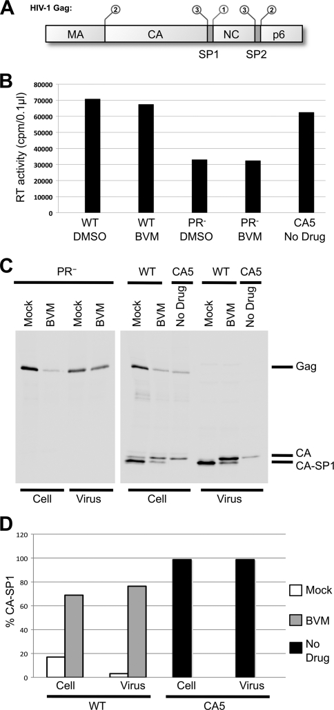
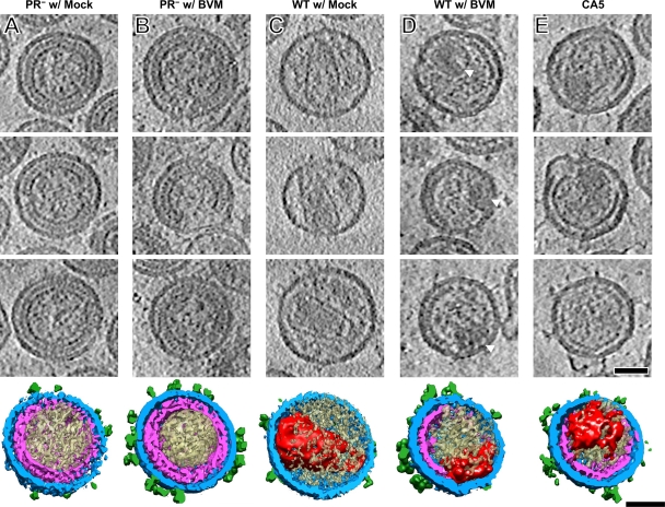
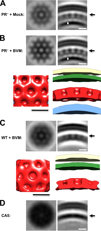
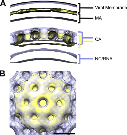
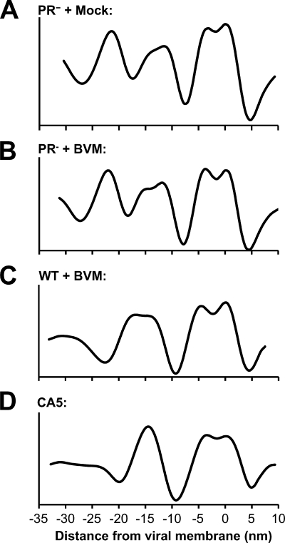
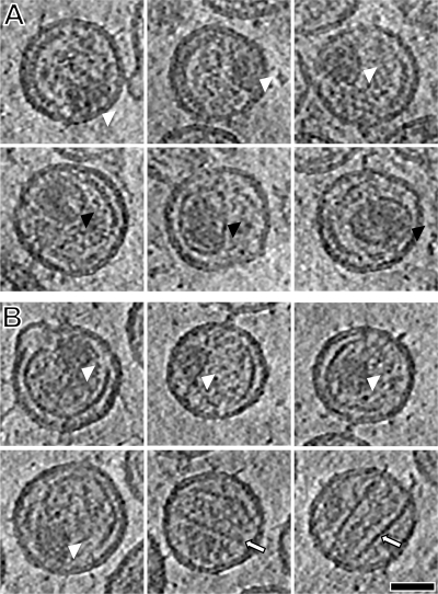
Maturation of nascent virions, a key step in retroviral replication, involves cleavage of the Gag polyprotein by the viral protease into its matrix (MA), capsid (CA), and nucleocapsid (NC) components and their subsequent reorganization. Bevirimat (BVM) defines a new class of antiviral drugs termed maturation inhibitors. BVM acts by blocking the final cleavage event in Gag processing, the separation of CA from its C-terminal spacer peptide 1 (SP1). Prior evidence suggests that BVM binds to Gag assembled in immature virions, preventing the protease from accessing the CA-SP1 cleavage site. To investigate this hypothesis, we used cryo-electron tomography to examine the structures of (noninfectious) HIV-1 viral particles isolated from BVM-treated cells. We find that these particles contain an incomplete shell of density underlying the viral envelope, with a hexagonal honeycomb structure similar to the Gag lattice of immature HIV but lacking the innermost, NC-related, layer. We conclude that the shell represents a remnant of the immature Gag lattice that has been processed, except at the CA-SP1 sites, but has remained largely intact. We also compared BVM-treated particles with virions formed by the mutant CA5, in which cleavage between CA and SP1 is also blocked. Here, we find a thinner CA-related shell with no visible evidence of honeycomb organization, indicative of an altered conformation and further suggesting that binding of BVM stabilizes the immature lattice. In both cases, the observed failure to assemble mature capsids correlates with the loss of infectivity.


Figures

FIG. 1.
FIG. 1.
(A) Domain organization and processing program of HIV-1 Gag. The functional domains MA, CA, NC, and p6, plus spacer peptides SP1 and SP2, are indicated. The SP1-NC cleavage occurs first (1), followed by separation of MA from CA and NC-SP2 from p6 (2). The final cleavage events separate NC from SP2 and CA from SP1 (3). (B) Virus particle production was measured by monitoring RT activity in the concentrated virus pellets. RT activity is 2-fold lower in PR samples, but sufficient quantities of particles to perform cryo-ET were obtained for all samples. (C and D) Detection of virus-associated proteins. HeLa cells transfected with WT pNL4-3 or derivatives and cultured in either the absence of drug or the presence of 4 μg/ml BVM or the equivalent concentration of DMSO were metabolically labeled for 3.5 h with [35S]Met-Cys, and released virions were pelleted by ultracentrifugation. Virus lysates were immunoprecipitated with anti-HIV-Ig, and processing of CA-SP1 to CA was analyzed by SDS-PAGE and fluorography (C) followed by phosphorimager analysis to quantify the percentage of CA-SP1 relative to total CA-SP1 plus CA (D).
FIG. 2.
FIG. 2.
Cryo-electron tomography of five HIV-1-related particles: pNL4-3 PR (immature) with mock treatment (A), pNL4-3 PR treated with 4 μg/ml BVM (B), pNL4-3 WT with mock treatment (C), pNL4-3 WT treated with 4 μg/ml BVM (D), and pNL4-3 CA5 (E). The three upper panels in each column show central sections, 0.78 nm thick, through three representative particles from that sample. Below, a segmented surface rendering of the particle in the bottom section is shown. The glycoprotein spikes are shown in green, the membrane plus MA layer is in blue, Gag-related shells are in magenta, core structures are in red, and other internal density is shown in beige. Scale bar, 50 nm.
FIG. 3.
FIG. 3.
Structural analysis of immature HIV-1 virions, BVM-treated virions, and CA5 virions. In each case, subtomogram averaging was performed to enhance the structures present in the outer density layers. (A and B) Immature virions from the protease-defective clone pNL4-3 PR. The samples were produced in the presence of DMSO (mock treatment; A) or BVM (B). White arrows indicate the putative helical bundles formed by SP1 connecting CA and NC/RNA densities. (C) Maturation-inhibited virions produced by WT pNL4-3 produced in the presence of 4 μg/ml BVM. (D) Virions from the mutant clone pNL4-3 CA5 produced in the absence of BVM. In the upper portions of panels A to C, two views of the averaged density map are shown: at left is an in-plane section, 0.78 nm thick, at the level marked with a long arrowhead in the CA density layer; at right is a radial section of similar thickness. In the lower portions of panels B and C are isodensity surface renderings of the respective structures. The viral membrane is shown in beige, the MA layer is in green, the CA layer is in red, and the NC/RNA layer, when visualized, is in blue. CA5 particles display no evidence of a lattice in the averaged in-plane section, from which we conclude that if such a lattice is present, the contrast and/or resolution of this analysis is not sufficient to detect it. Scale bar, 10 nm.
FIG. 4.
FIG. 4.
Comparison of immature and BVM-treated lattices. Average density maps from PR virus treated with 4 μg/ml BVM (blue; mesh) and WT virus treated with 4 μg/ml BVM (beige; surface) were aligned and compared. (A) Radial view of density maps aligned with viral membrane present. Density layers are labeled with the corresponding viral structures. The membrane and MA layers are essentially superposable, whereas the CA density layer has shifted inwards in the BVM-treated particle following PR cleavage. (B) Top view of CA lattice. The aligned hexagonal lattices are shown superimposed in this view. Scale bar, 10 nm.
FIG. 5.
FIG. 5.
Comparison of radial density profiles of immature virions, BVM-treated virions, and CA5 virions. Radial density profiles were calculated from subtomogram averages of immature virions produced in the presence of DMSO (A), immature virions produced in the presence of BVM (B), WT virions produced in the presence of BVM (C), and CA5 particles produced in the absence of BVM (D). Profiles have been aligned based on the position of the viral membrane.
FIG. 6.
FIG. 6.
Gallery of core structures in BVM-treated and CA5 particles: pNL4-3 WT with BVM treatment (A) and pNL4-3 CA5 (B). Central sections, 0.78 nm thick, through eight representative particles are shown for each sample. Core-like features are indicated by arrowheads: white, small, electron-dense material with no shell; black, defined shell containing electron-dense material; outlined arrows, incomplete tubes and cones. Scale bar, 50 nm.

References

    1. Adachi, A., et al. 1986. Production of acquired immunodeficiency syndrome-associated retrovirus in human and nonhuman cells transfected with an infectious molecular clone. J. Virol. 59:284-291. - PMC - PubMed
    1. Adamson, C. S., et al. 2006. In vitro resistance to the human immunodeficiency virus type 1 maturation inhibitor PA-457 (bevirimat). J. Virol. 80:10957-10971. - PMC - PubMed
    1. Adamson, C. S., and E. O. Freed. 2007. Human immunodeficiency virus type 1 assembly, release, and maturation. Adv. Pharmacol. 55:347-387. - PubMed
    1. Adamson, C. S., and E. O. Freed. 2008. Recent progress in antiretrovirals—lessons from resistance. Drug Discov. Today 13:424-432. - PMC - PubMed
    1. Adamson, C. S., M. Sakalian, K. Salzwedel, and E. O. Freed. 2010. Polymorphisms in Gag spacer peptide 1 confer varying levels of resistance to the HIV-1 maturation inhibitor bevirimat. Retrovirology 7:36. - PMC - PubMed

Publication types

MeSH terms