. 2011 Feb;85(4):1541-53.
doi: 10.1128/JVI.01790-10. Epub 2010 Nov 24.

Cell type-specific proteasomal processing of HIV-1 Gag-p24 results in an altered epitope repertoire

Affiliations

Cell type-specific proteasomal processing of HIV-1 Gag-p24 results in an altered epitope repertoire

Nicholas J Steers et al. J Virol. 2011 Feb.

Abstract

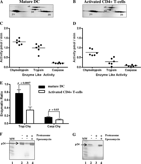
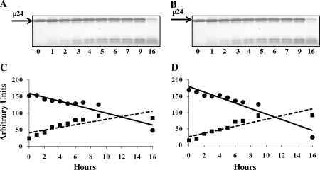
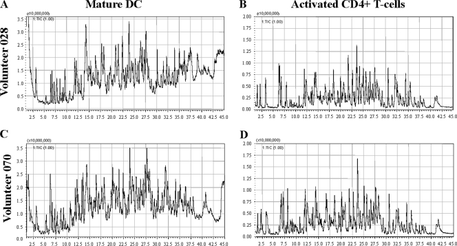
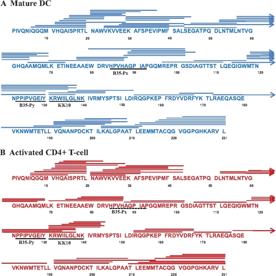
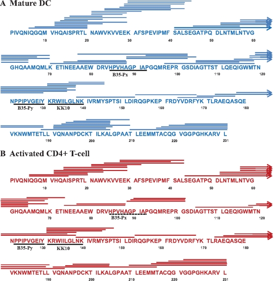
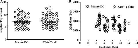
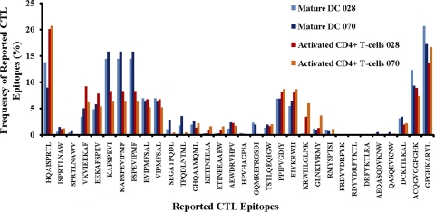
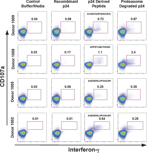
Proteasomes are critical for the processing of antigens for presentation through the major histocompatibility complex (MHC) class I pathway. HIV-1 Gag protein is a component of several experimental HIV-1 vaccines. Therefore, understanding the processing of HIV-1 Gag protein and the resulting epitope repertoire is essential. Purified proteasomes from mature dendritic cells (DC) and activated CD4(+) T cells from the same volunteer were used to cleave full-length Gag-p24 protein, and the resulting peptide fragments were identified by mass spectrometry. Distinct proteasomal degradation patterns and peptide fragments were unique to either mature DC or activated CD4(+) T cells. Almost half of the peptides generated were cell type specific. Two additional differences were observed in the peptides identified from the two cell types. These were in the HLA-B35-Px epitope and the HLA-B27-KK10 epitope. These epitopes have been linked to HIV-1 disease progression. Our results suggest that the source of generation of precursor MHC class I epitopes may be a critical factor for the induction of relevant epitope-specific cytotoxic T cells.


Figures

FIG. 1.
FIG. 1.
Compositions and enzyme activities of proteasomes isolated from mature human DC and activated CD4+ T cells. Primary DC and CD4+ T cells were isolated from PBMCs derived from leucopacks of volunteer 070. The purified proteasomes from mature DC (A) and activated CD4+ T cells (B) were analyzed by 2-D IEF followed by Western blotting with antibodies specific for β1i, β2i, β5i, PA28α, and PA28β. (C, D) Proteasomes isolated from both mature DC and activated CD4+ T cells were characterized as i20s immunoproteasomes containing the β1i, β2i, and β5i subunits. Proteolytic activities of the purified proteasomes were measured to ensure that they retained their functional activities. The enzymatic profiles of mature DC (C) and activated CD4+ T cells (D) showed high chymotrypsin and trypsin-like activities, with a much lower caspase-like activity. (E) The ratios of the enzymatic activities of the purified proteasomes from mature DC and activated CD4+ T cells were plotted based on the data shown in panels C and D. The trypsin (Tryp)/chymotrypsin (Chy) and caspase (Casp)/chymotrypsin ratios were determined for proteasomes isolated from each cell type. The specificities of the proteolytic cleavage of Gag-p24 (subtype B) in purified proteasomes from mature DC (F) and activated CD4+ T cells (G) were demonstrated by the addition of epoxomycin, an irreversible proteasome inhibitor that prevented proteasomal cleavage of Gag-p24. Samples were run on a 4-to-20% gradient Tris-glycine polyacrylamide gel. MW, molecular weight markers.
FIG. 2.
FIG. 2.
Time course of the proteasomal cleavage of Gag-p24. (A, B) Gag-p24 (10 μg) was incubated with purified proteasomes (2 μg) from mature DC (A) and activated CD4+ T cells (B) for the indicated time points (0 to 16 h) and then frozen at −80°C to terminate the reaction. (C, D) Samples were run on a 4 to 20% Tris-glycine gel and stained with Pierce blue to determine the intensities of the remaining Gag-p24 and the proteolytically cleaved products. The gels were dried and scanned, and the intensities were analyzed using ImageJ Software to quantify the cleaved and the accumulated Gag-p24 degradation products. There was a reciprocal correlation between the proteolytic cleavage (•) of Gag-p24 and the accumulation (▪) of the degradation products.
FIG. 3.
FIG. 3.
Total ion chromatogram (TIC) profiles following the proteasomal degradation of Gag-p24. Gag-p24 (25 μg) was cleaved using purified proteasomes (5 μg) isolated from mature DC (A and C) and primary CD4+ T cells (B and D) from volunteers 028 and 070, respectively. The degradation mixture was analyzed using a SHIM-PACK XR-ODS II 2.0-mm by 150-mm column on a UFLC. The peptides were eluted using a 3 to 35% acetonitrile gradient containing 0.1% formic acid. The graphical data represent the total ion counts (y axis) over a 45-min time period (x axis). The peptides were then directly analyzed on a LCMS-IT-TOF mass spectrometer.
FIG. 4.
FIG. 4.
Proteasomal cleavage maps of Gag-p24. The proteasomal peptide fragments were separated by UFLC, analyzed on an LCMS-IT-TOF mass spectrometer, and then identified using the Mascot database. The Gag-p24 peptides generated from proteasomes isolated from mature DC (A) and activated CD4+ T cells (B) from volunteer 028 were identified. The lines above the sequence represent the identified peptide fragments. The arrows above the Gag-p24 sequences denote the continuation of the peptide fragments. The solid lines and dashed lines below the sequences denote the generation and absence of specific epitopes, respectively. The KK10 epitope and the B35-Px epitope are differentially produced in the two cell types. The frequencies of peptides containing the KK10 epitope for the activated CD4+ T cells were 2.48% (KRWIILGLNKIVRM) and 0.75% (KRWIILGLNKIVRMY), compared to 0.2% (KRWIILGLNKIVRM) for mature DC. The B35-Px epitope was absent in activated CD4+ T cells. The frequency of the fragment containing this epitope (WDRVHPVHAGPIAPGQMREPRG) in mature DC was 0.3%.
FIG. 5.
FIG. 5.
Proteasomal cleavage maps of Gag-p24. The proteasomal peptide fragments were separated by UFLC, analyzed on an LCMS-IT-TOF mass spectrometer, and then identified using the Mascot database. The Gag-p24 peptides generated from proteasomes isolated from mature DC (A) and activated CD4+ T cells (B) from volunteer 070 were identified. The lines above the sequences represent the identified peptide fragments. The arrows above the Gag-p24 sequences denote the continuation of the peptide fragments. The solid lines and dashed lines below the sequences denote the generation and absence of specific epitopes, respectively. The KK10 epitope and the B35-Px epitope are differentially produced in the two cell types. The frequencies of peptides containing the KK10 epitope for the activated CD4+ T cells were 0.13% (KRWIILGLNKI), 3.25% (KRWIILGLNKIVRM), and 2.57% (KRWIILGLNKIVRMY), compared to 0.2% (KRWIILGLNKIVRM) for the mature DC. The B35-Px epitope was absent in the activated CD4+ T cells, and the frequency for the fragment(s) containing this epitope (AAEWDRVHPVHAGPIAPGQMREPRG) in mature DC was 0.32%.
FIG. 6.
FIG. 6.
Biochemical characteristics of the identified proteasomal degradation products of Gag-p24. (A) The peptides identified from mature DC and activated CD4+ T cells from volunteer 070 shown in Fig. 5A and B, respectively, were plotted to compare the lengths of the peptide sequences produced by proteasomes from the two cell types. (B) The molecular masses and the isoelectric points of the peptides were also analyzed. No significant differences in the amino acid (aa) lengths (A), pIs, or molecular masses (B) of the peptides produced from the cleavage products of proteasomes from either mature DC or activated CD4+ T cells were observed.
FIG. 7.
FIG. 7.
Graphical representation of Gag-p24 proteasomal digests. The relative peak areas of all the peptides identified from mature DC (Fig. 4A and 5A) and activated CD4+ T cells (Fig. 4B and 5B) from two volunteers (028 and 070) were determined by peak integration from the extracted ion chromatograms. The relative frequencies of the identified peptides from the proteasomal digests of mature DC (blue columns) and activated CD4+ T cells (red columns) from each of the volunteers were calculated using ion chromatograms relative to each peptide fragment based on the area under the fraction of each peptide peak. This is plotted as the percentage of each peptide relative to the total number of identified peptides from each cell type. The data demonstrate both similarities and differences between the degradation products from the two cell types.
FIG. 8.
FIG. 8.
Relative frequencies of the best-defined CTL epitopes derived from the peptides identified from the proteasomal degradation of Gag-p24. The relative frequency of each of the CTL epitopes from mature DC (blue columns) and activated CD4+ T cells (red columns) from volunteers 028 and 070 was calculated using the frequency of peptides identified in Fig. 7 that contained the reported CTL epitopes.
FIG. 9.
FIG. 9.
Antigen presentation of Gag-p24 proteasomal digests. Proteasomal digests of Gag-p24 from activated CD4+ T cells were incubated with PBMCs from four different HIV-1-positive individuals. PBMCs were incubated with Gag-p24 protein, 15-mer synthetic peptides derived from Gag-p24, SEB (data not shown), and media containing proteasomal digestion buffer. CD3+ CD8+ T cells were gated from PBMCs and then analyzed for the induction of IFN-γ and CD107a by ICS. In cells from all four volunteers, there was no induction of CD8+ T-cell-specific IFN-γ or CD107a in the presence of media (first column) or following incubation with Gag-p24 (second column). In contrast, the proteasome-derived Gag-p24 peptides induced a 10- to 80-fold induction of CD8+ T cells specific for IFN-γ and CD107a (last column) compared to that in the buffer/medium-alone samples, and this induction was similar to or higher than the results obtained with synthetic 15-mer peptides containing CTL epitopes of Gag-p24 (third column).

Similar articles

Cited by

References

    1. Ackerman, A. L., A. Giodini, and P. Cresswell. 2006. A role for the endoplasmic reticulum protein retrotranslocation machinery during crosspresentation by dendritic cells. Immunity 25:607-617. - PubMed
    1. Altfeld, M., et al. 2003. Influence of HLA-B57 on clinical presentation and viral control during acute HIV-1 infection. AIDS 17:2581-2591. - PubMed
    1. Bansal, A., et al. 2007. Immunological control of chronic HIV-1 infection: HLA-mediated immune function and viral evolution in adolescents. AIDS 21:2387-2397. - PMC - PubMed
    1. Barouch, D. H., et al. 2010. Mosaic HIV-1 vaccines expand the breadth and depth of cellular immune responses in rhesus monkeys. Nat. Med. 16:319-323. - PMC - PubMed
    1. Basler, M., N. Youhnovski, M. Van Den Broek, M. Przybylski, and M. Groettrup. 2004. Immunoproteasomes down-regulate presentation of a subdominant T cell epitope from lymphocytic choriomeningitis virus. J. Immunol. 173:3925-3934. - PubMed

Publication types

MeSH terms