. 2010 Dec 14;107(50):21671-6.
doi: 10.1073/pnas.1016233107. Epub 2010 Nov 24.

Target-seeking antifibrotic compound enhances wound healing and suppresses scar formation in mice

Affiliations

Target-seeking antifibrotic compound enhances wound healing and suppresses scar formation in mice

Tero A H Järvinen et al. Proc Natl Acad Sci U S A. .

Abstract

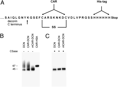
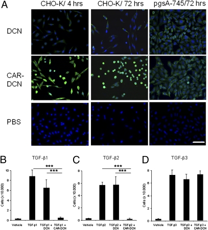
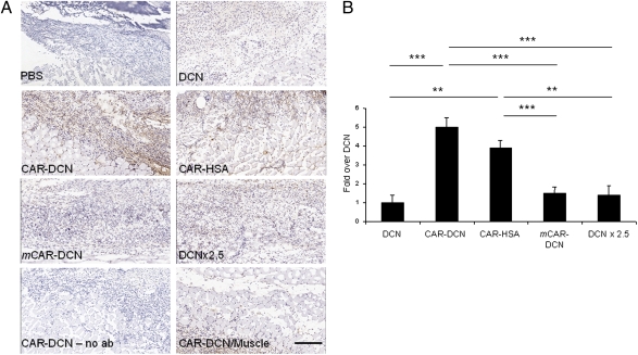
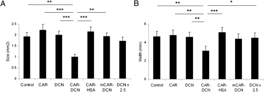
Permanent scars form upon healing of tissue injuries such as those caused by ischemia (myocardial infarction, stroke), trauma, surgery, and inflammation. Current options in reducing scar formation are limited to local intervention. We have designed a systemically administered, target-seeking biotherapeutic for scar prevention. It consists of a vascular targeting peptide that specifically recognizes angiogenic blood vessels and extravasates into sites of injury, fused with a therapeutic molecule, decorin. Decorin prevents tissue fibrosis and promotes tissue regeneration by inhibiting TGF-β activity and by other regulatory activities. The decorin-targeting peptide fusion protein had substantially increased neutralizing activity against TGF-β1 in vitro compared with untargeted decorin. In vivo, the fusion protein selectively accumulated in wounds, and promoted wound healing and suppressed scar formation at doses where nontargeted decorin was inactive. These results show that selective targeting yields a tissue-healing and scar-reducing compound with enhanced specificity and potency. This approach may help make reducing scar formation by systemic drug delivery a feasible option for surgery and for the treatment of pathological processes in which scar formation is a problem.


Conflict of interest statement

The authors declare no conflict of interest.

Figures

Fig. 1.
Fig. 1.
Cloning and production of recombinant fusion proteins. (A) Schematic representation of the CAR–decorin fusion protein showing the insertion of the CAR peptide sequence and a polyhistidine-tag C-terminal of full-length decorin. (B) Gel electrophoretic analysis of recombinant decorins and CAR–HSA. The recombinant proteins were expressed in mammalian cells, purified on a Ni-column, separated on gradient SDS/PAGE gels, and detected with a monoclonal anti–6-histidine tag antibody. The decorins migrate as sharp bands at 45 kDa with a smear above the sharp band. (C) Digestion of the recombinant decorins with chondroitinase ABC before electrophoresis removed the smear. Thus, the sharp bands correspond to the core proteins and the smear is caused by the chondroitin sulfate chain attached to most of the decorin molecules (7, 8, 22, 23).
Fig. 2.
Fig. 2.
Cell binding and inhibition of cell spreading and TGF-β–dependent cell proliferation by decorins. (A) The culture media of CHO-K and pgsA-745 cells were supplemented with 0.3 μg/mL of decorins daily for 4 h or 3 d. After washing, the decorins were detected with anti–his-tag antibody and FITC-conjugated secondary antibody (green). The CAR-targeted decorin bound to CHO-K cells more (4 h) and inhibited cell spreading more strongly (3 d) than unmodified decorin, whereas the two decorins show identical low binding to mutant pgsA-745 cells that lack the receptor for the CAR peptide (19). Cell nuclei were stained by DAPI (blue). (Scale bar, 50 μm.) (B) CHO-K cells were grown in the presence of TGF-β1, (C) TGF-β2, or (D) TGF-β3 (all at 7.5 ng/mL) and 0.3 μg/mL of decorins. Half of the medium was changed daily for 4 d (7). Error bars represent mean ± SD. Two to four separate experiments were performed in triplicate. CAR-targeted decorin was particularly potent in inhibiting TGF-β1– and -β2–driven cell proliferation (B and C, ***P ≤ 0.001 both compared with control and decorin; ANOVA).
Fig. 3.
Fig. 3.
Homing of CAR–decorin in wound tissue. Mice with full thickness skin wounds received an i.v. injection of His-tagged fusion proteins on day 5 after wounding. After 4 h, the location of decorin was determined with an anti–His-tag antibody (brown). (A) The wounds of mice injected with nontargeted decorin (DCN) or mCAR–DCN were weakly positive, whereas strong staining was observed in CAR–DCN wounds. The accumulation of CAR–HSA in the wounds confirmed the ability of the CAR peptide to enhance targeting to the wounds. No staining was observed in normal skeletal muscle underlying the skin wounds of mice treated with any of the decorins (shown for CAR–DCN; CAR–DCN/muscle). No staining was seen in wound tissue when class-matched mouse IgG was substituted for the anti–6-histidine tag antibody (no ab). (Scale bar, 200 μm.) (B) Quantitative analysis of homing in skin wounds. The statistical significance was examined with ANOVA; *P < 0.05, **P ≤ 0.01, ***P < 0.001, n = 16 per group. The results are expressed as mean ± SD.
Fig. 4.
Fig. 4.
Reduced scar formation during wound healing in mice treated with CAR–decorin. Mice with full thickness skin wounds received daily i.v. injections as indicated from day 3 after the wounding until day 14. Scars were harvested on day 21, and the cross-sectional area (A) and the width (diameter) (B) of the scars were quantified by examining two microscopic sections from each wound. The results are expressed as the average of the two values. There were seven animals, each with four wounds, in every treatment group. *P < 0.05, **P < 0.01, ***P < 0.001; ANOVA. The results are expressed as mean ± SEM, n = 28.
Fig. 5.
Fig. 5.
α-SMA–positive fibroblasts (myofibroblasts) and connective tissue growth factor (CCN2/CTGF) in wounds treated with targeted decorin. Representative sections from wounds collected on day 10 after wounding are shown for α-SMA–positive myofibroblasts (A) and for CCN2/CTGF (B). A wound from a decorin-treated mouse was stained with class-matched mouse IgG as a specificity control (no ab). The wounds of mice treated with CAR–DCN showed diminished myofibroblast reaction and CCN2/CTGF protein expression in their skin wounds. The arrows indicate α-SMA–positive smooth muscle cells in the walls of blood vessels in CAR–DCN treated wound. Quantitative analysis of α-SMA–positive myofibroblasts (C) and CCN2/CTGF protein expression (D) in skin wounds at day 10. The statistical significance was examined with ANOVA, *P < 0.05, **P ≤ 0.01, ***P ≤ 0.001. The results are expressed as mean ± SEM, n = 16. (Scale bars, 150 μm.) (C) PBS/BSA control treatment; CAR refers to the CAR peptide.

References

    1. Aarabi S, Longaker MT, Gurtner GC. Hypertrophic scar formation following burns and trauma: New approaches to treatment. PLoS Med. 2007;4:e234. - PMC - PubMed
    1. Shaw TJ, Martin P. Wound repair at a glance. J Cell Sci. 2009;122:3209–3213. - PMC - PubMed
    1. Gurtner GC, Werner S, Barrandon Y, Longaker MT. Wound repair and regeneration. Nature. 2008;453:314–321. - PubMed
    1. Border WA, Ruoslahti E. Transforming growth factor-β in disease: The dark side of tissue repair. J Clin Invest. 1992;90:1–7. - PMC - PubMed
    1. Brunner G, Blakytny R. Extracellular regulation of TGF-β activity in wound repair: Growth factor latency as a sensor mechanism for injury. Thromb Haemost. 2004;92:253–261. - PubMed

Publication types

MeSH terms