Comparative Study
. 2010 Nov 24;30(47):15801-10.
doi: 10.1523/JNEUROSCI.1569-10.2010.

Task-dependent changes in short-term memory in the prefrontal cortex

Affiliations
Comparative Study

Task-dependent changes in short-term memory in the prefrontal cortex

Melissa R Warden et al. J Neurosci. .

Abstract

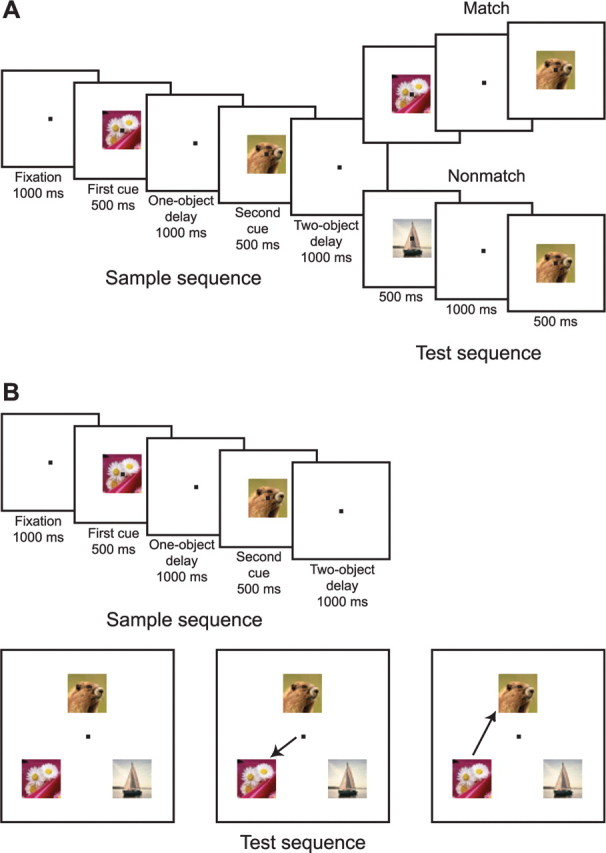
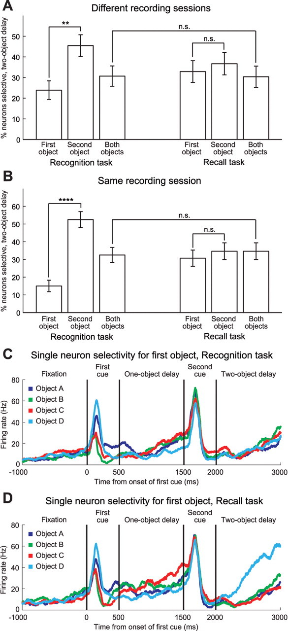
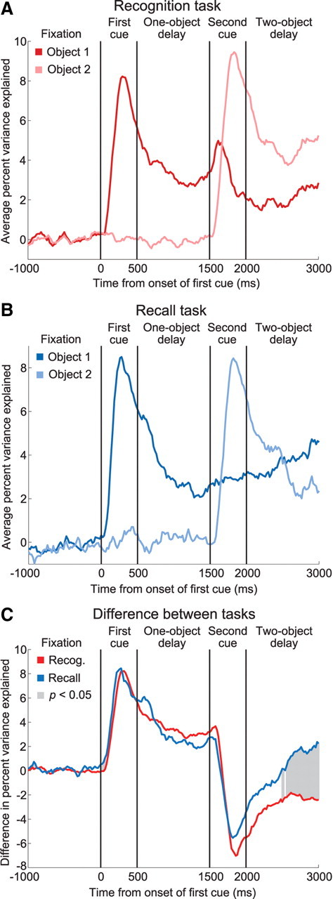
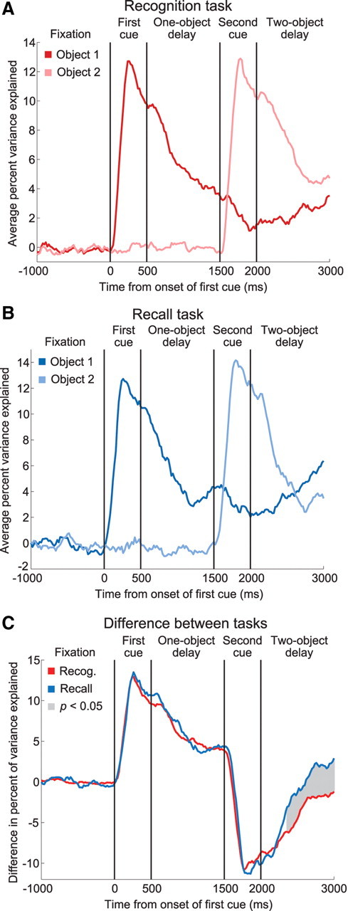
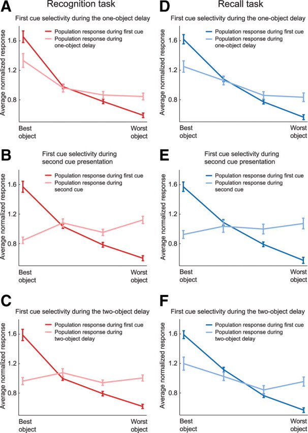
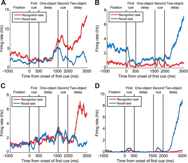
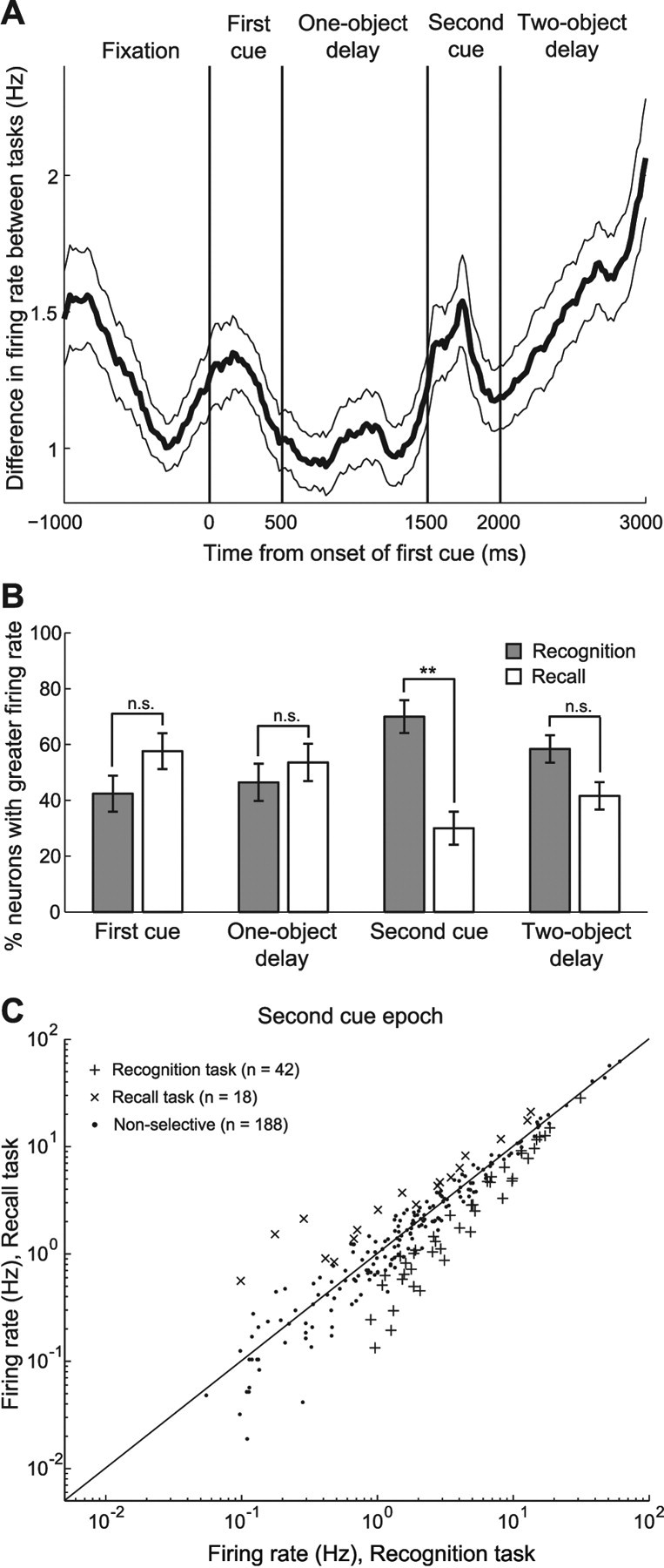
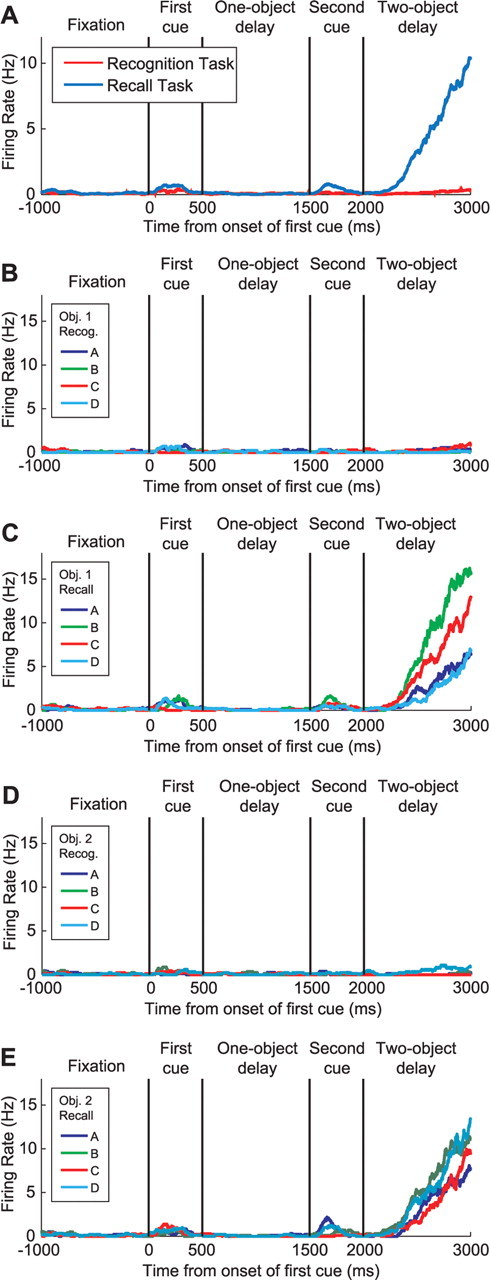
The prefrontal cortex (PFC) is important for flexible, context-dependent behavioral control. It also plays a critical role in short-term memory maintenance. Though many studies have investigated these functions independently, it is unclear how these two very different processes are realized by a single brain area. To address this, we trained two monkeys on two variants of an object sequence memory task. These tasks had the same memory requirements but differed in how information was read out and used. For the "recognition" task, the monkeys had to remember two sequentially presented objects and then release a bar when a matching sequence was recognized. For the "recall" task, the monkeys had to remember the same sequence of objects but were instead required to recall the sequence and reproduce it with saccadic eye movements when presented with an array of objects. After training, we recorded the activity of PFC neurons during task performance. We recorded 222 neurons during the recognition task, 177 neurons during the recall task, and 248 neurons during the switching task (interleaved blocks of recognition and recall). Task context had a profound influence on neural selectivity for objects. During the recall task, the first object was encoded more strongly than the second object, while during the recognition task, the second object was encoded more strongly. In addition, most of the neurons encoded both the task and the objects, evidence for a single population responsible for these two critical prefrontal functions.


Figures

Figure 1.
Figure 1.
Behavioral tasks. A, For the recognition (bar-release) task, each trial began when the monkeys grasped a bar and achieved central fixation. The first sample object was followed by a brief delay, then the second sample object, then another delay. This sample phase of the task was immediately followed by a test sequence with an identical temporal structure. The test sequence was either a match to the sample, in which case the monkeys were required to release the bar, or a nonmatch, in which case the monkeys were required to release the bar during a subsequent match sequence. B, The sample phase of the recall (saccade choice) task was identical to that of the recognition task. However, it was instead followed by an array of three test objects. The monkeys were required to make a correct sequence of saccades to the two objects seen during the sample phase. In the switching task, the monkeys performed interleaved blocks of both the recognition trials and the recall trials during the same recording session.
Figure 2.
Figure 2.
Strength of object representations. A, The proportion of the neural population representing only the first object, only the second object, or both objects during the two-object memory delay. In the recognition task, twice as many neurons represented the second object as the first object. In the recall task, the first and the second objects were represented equally. In both tasks, about one-third of the population represented both objects. n.s. indicates not significant, **p < 0.01, and ****p < 0.0001. Error bars indicate SE. B, The same analysis, with the same conclusions, repeated for the switching task. C, A single neuron recorded during the recognition task, trials grouped according to which object was used as the first cue. This neuron showed little selectivity for the first object at any point during the trial. D, The same neuron recorded during the recall task, trials again grouped by the first object. The same neuron showed strong selectivity for the first object during the two-object delay when the monkey performed the recall task.
Figure 3.
Figure 3.
Relative object strengths as a function of time in both tasks, separate recording sessions. A, The percentage variance explained by the first or the second object during the recognition task, averaged across the population of neurons. During the two-object delay period, the most recently seen object has a stronger representation. B, The same analysis during the recall task. Now, the most recent object has a weaker representation during the two-object delay. C, The object 1 curve minus the object 2 curve for both tasks. The relative strengths of the object representations during the two-object delay depend on which task the monkeys are performing. The shaded gray area indicates a significant difference in relative strengths (two-sample t test, p < 0.05).
Figure 4.
Figure 4.
Relative object strengths during interleaved blocks of the recognition and recall tasks. A, The percentage variance explained by the first or the second object during the recognition task, averaged across the population of neurons. Again, these neurons show a stronger representation of the most recently seen object. B, The same analysis during the recall task. As seen during the separate recording sessions, the neurons show a weaker representation of the most recent object during this task. C, The object 1 curve minus the object 2 curve for both tasks. Even though the monkeys were switching between the tasks frequently, the task had a significant effect on the relative strengths of the objects.
Figure 5.
Figure 5.
A–C, Recognition task. A, Normalized response of each neuron to the first object during both the first cue period (red) and the one-object delay period (pink), averaged across the population of selective neurons, ordered best to worst object as defined by the response during the first cue period. The population maintains its object preferences during the one-object delay period. B, Response to the first object during both the first cue period (red) and the second cue period (pink). The inversion of the slope shows that many neurons have changed preferred first objects. C, Response to the first object during both the first cue period (red) and the two-object delay period (pink). The flattening of this curve shows that many neurons have changed preferred first objects, but, when averaged together, the responses produce a flat line. D–F, Recall task. The same analysis as in A–C is shown, using data obtained during the recall task. D, Response to the first object during the first cue period (dark blue) is similar to the response to this object during the one-object delay (light blue). E, The response to the first object during the second cue period is flattened, indicating a change in selectivity. F, The response to the first object during the two-object delay period has a positive slope again, reflecting the reacquisition of initial object preferences as seen during cue presentation.
Figure 6.
Figure 6.
Four individual neurons recorded during the switching task show task-dependent differences in firing rate. A, C, Two neurons that have a higher firing rate during the recognition task. B, D, Two neurons that have a higher firing rate during the recall task.
Figure 7.
Figure 7.
Task-dependent differences in firing rate. A, Time course of the difference in firing rate between tasks averaged across the population of neurons, ±SE. B, Relative proportions of neurons more responsive during the recognition or recall task for each task epoch. n.s. indicates not significant, and ** indicates p < 0.01, one-proportion Z test. Error bars indicate SE. C, Individual neurons showed task-dependent differences in firing rate. Each point in this figure represents the activity of one neuron during the second cue epoch. Average firing rate during the recognition task is plotted against average firing rate during the recall task. Neurons that show significantly different firing rates between tasks are plotted using + or ×. The data are plotted logarithmically due to the spread of the data and the large number of points at low firing rates.
Figure 8.
Figure 8.
Anatomical locations of recording sites and selective neurons in both monkeys during the switching task. ×, ○, Recording sites at which neurons selective for objects and/or task during the two-object delay period were found, respectively. Black dots, Locations at which neurons were recorded but no selective neurons were encountered. Multiple neurons were recorded at many locations.
Figure 9.
Figure 9.
A single neuron selective for both task and object. A, A neuron that fires more strongly during the recall task. B, Object selectivity of the same neuron during the recognition task. The trials are grouped according to which object was presented as the first cue. C, Object selectivity of the same neuron during the recall task. The trials are again grouped according to which object was presented as the first cue. D, Selectivity for the second object during the recognition task. E, Selectivity for the second object during the recall task.

References

    1. Asaad WF, Rainer G, Miller EK. Task-specific neural activity in the primate prefrontal cortex. J Neurophysiol. 2000;84:451–459. - PubMed
    1. Averbeck BB, Chafee MV, Crowe DA, Georgopoulos AP. Parallel processing of serial movements in prefrontal cortex. Proc Natl Acad Sci U S A. 2002;99:13172–13177. - PMC - PubMed
    1. Brown J. Some tests of the decay theory of immediate memory. Q J Exp Psychol. 1958;10:12–21.
    1. Cabeza R, Kapur S, Craik FIM, McIntosh AR, Houle S, Tulving E. Functional neuroanatomy of recall and recognition: a PET study of episodic memory. J Cogn Neurosci. 1997;9:254–265. - PubMed
    1. Delbecq-Derouesné J, Beauvois MF, Shallice T. Preserved recall versus impaired recognition. A case study. Brain. 1990;113:1045–1074. - PubMed

Publication types

LinkOut - more resources