Comparative Study
. 2010 Nov 24;30(47):15821-33.
doi: 10.1523/JNEUROSCI.3448-10.2010.

Orchestration of stepwise synaptic growth by K+ and Ca2+ channels in Drosophila

Affiliations
Comparative Study

Orchestration of stepwise synaptic growth by K+ and Ca2+ channels in Drosophila

Jihye Lee et al. J Neurosci. .

Abstract

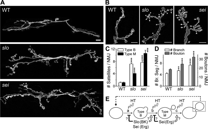
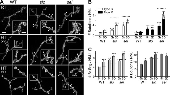
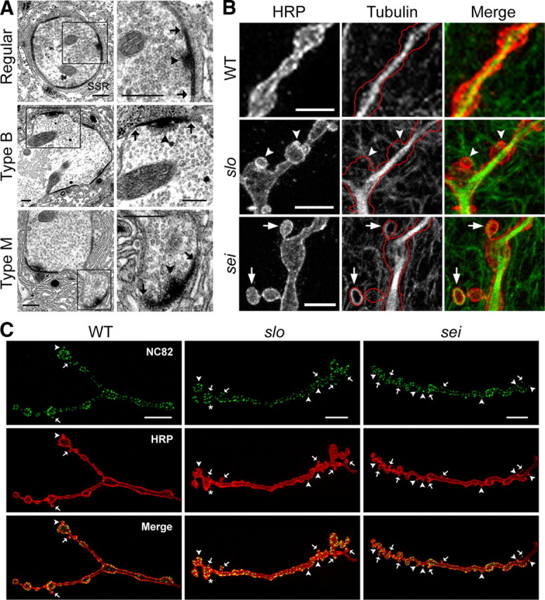
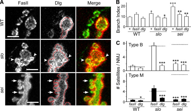
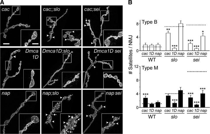
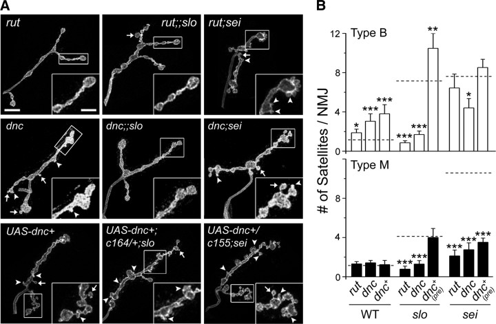
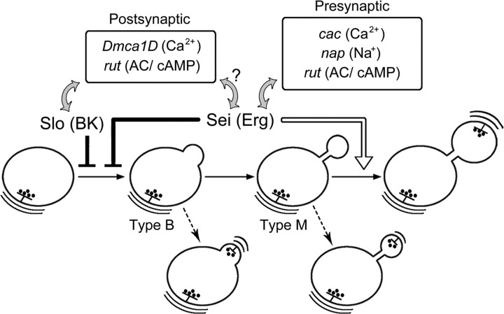
Synapse formation is tightly associated with neuronal excitability. We found striking synaptic overgrowth caused by Drosophila K(+)-channel mutations of the seizure and slowpoke genes, encoding Erg and Ca(2+)-activated large-conductance (BK) channels, respectively. These mutants display two distinct patterns of "satellite" budding from larval motor terminus synaptic boutons. Double-mutant analysis indicates that BK and Erg K(+) channels interact with separate sets of synaptic proteins to affect distinct growth steps. Post-synaptic L-type Ca(2+) channels, Dmca1D, and PSD-95-like scaffold protein, Discs large, are required for satellite budding induced by slowpoke and seizure mutations. Pre-synaptic cacophony Ca(2+) channels and the NCAM-like adhesion molecule, Fasciclin II, take part in a maturation step that is partially arrested by seizure mutations. Importantly, slowpoke and seizure satellites were both suppressed by rutabaga mutations that disrupt Ca(2+)/CaM-dependent adenylyl cyclase, demonstrating a convergence of K(+) channels of different functional categories in regulation of excitability-dependent Ca(2+) influx for triggering cAMP-mediated growth plasticity.


Figures

Figure 1.
Figure 1.
Aberrant synaptic growth induced by slo and sei K+-channel mutations. A, B, Representative anti-HRP immunostaining of WT, slo, and sei larval NMJs in muscles 6 and 7 (A) and muscle 4 (M4; type Ib only; B) of abdominal segments 3 or 4. Note the more extensive branching at slo and sei NMJs (A) and small “satellite” boutons in these mutants budding from larger, primary synaptic boutons either with (type M, arrows) or without (type B, arrowheads) a clear constriction (B). Fluorescent micrographs in this and subsequent figures are collapsed confocal Z-stacks. Scale bars: A, 10 μm; B, 5 μm. C, D, Pooled data are shown for the numbers of types B and M satellites (C) and of mature primary boutons and terminal branches [branch segments (Br. Seg)] (D) per type Ib M4 NMJ. The number of NMJs (larvae) examined is as follows: 91 (30) for WT, 107 (34) for slo, and 38 (11) for sei. **p < 0.01; ***p < 0.001 (one-way ANOVA). E, Model of sequential synaptic growth process, from initial budding (type B satellites; step a) to maturation into type M satellites (step b) and formation of primary boutons (step c). Open arrows and closed bars indicate the alternative possibilities of promoting or restraining actions, respectively, exerted by these K+ channels at individual steps that might explain the mutant phenotypes (see Results). Facilitated growth by HT treatment and reiteration of a growth cycle (dashed line) are indicated. Error bars indicate mean ± SEM.
Figure 2.
Figure 2.
Dynamic synaptic growth regulation in slo and sei mutants revealed by short- and long-term exposure to high rearing temperature. A, Representative type Ib NMJs in muscle 4 (M4) of WT, slo, and sei larvae reared at RT (top) and after short-term (5 h; middle) or long-term [5 d (5D); bottom] exposure to high temperature (29°C). Examples of types B (arrowheads) and M (arrows) satellites are indicated. Note branches consisting predominantly of type M satellites (asterisks) that are found only in long-term, HT-treated sei larvae. Scale bars: A, 10 μm; insets, 5 μm. B, C, Pooled data for the numbers of types B and M satellites (B), terminal branches, and mature primary boutons (C) at type Ib M4 NMJs are shown for short- and long-term HT-treated WT, slo, and sei larvae. The control values at RT (from Fig. 1 for each genotype) are indicated by dashed lines. The portions of HT-induced small type B or M satellites forming strings at sei NMJs are indicated as hatched bars. The numbers of NMJs (larvae) examined are as follows: after HT for 5 h, WT, 23 (7); slo, 26 (7); sei, 12 (3); after HT for 5 d, WT, 22 (6); slo, 35 (8); sei, 24 (6). *p < 0.05; **p < 0.01; ***p < 0.001 [t test for RT vs HT (5 h or 5 d) within each genotype]. Error bars indicate mean ± SEM.
Figure 3.
Figure 3.
Synaptic ultrastructure and microtubule networks in slo and sei satellites. A, Electron microscopy images of mature boutons and satellites found in slo and sei mutants. In contrast to smooth contour of a WT bouton (top), the contours of slo and sei boutons appear irregular because of the presence of types B (middle) and M (bottom) satellites. Note the presence of electron-dense membrane areas (demarcated by arrows) and T-bar specializations (arrowheads) associated with synaptic active zones, as well as synaptic vesicles, in both types of satellites. The boxed region in the left column is magnified on the right. B, Confocal images of type Ib NMJs in muscle 4 (M4) of WT, slo, and sei larvae displaying networks of microtubules revealed by α-tubulin immunoreactivity (middle). Pre-synaptic bouton contours (red lines) are traced from anti-HRP images (left) against α-tubulin images (middle). Note the lack of clear tubulin immunoreactivity within type B satellites (slo; arrowheads), in contrast to a distinct loop structure in type M satellites (sei; arrows). C, Confocal images of type Ib NMJs in M4 of WT, slo, and sei larvae displaying overall distributions of active zones visualized by NC82 antibody against Brp, a protein important for T-bar integrity at active zones (top). General morphology of each NMJ is revealed by double staining of HRP at the same NMJs (middle). Note clear NC82 signals in type B (arrowheads) and M (arrows) satellites with a rare exception (asterisk). Scale bars: A, 0.5 μm; B, 5 μm; C, 10 μm.
Figure 4.
Figure 4.
Distribution of Dlg scaffold and FasII adhesion proteins and suppression of slo and sei satellites by dlg and fasII mutations. A, Immunoreactivity against FasII (left) and Dlg (middle) at WT (types Ib and Is), slo, and sei (type Ib only) muscle 4 NMJs. The contour of boutons outlined by FasII immunoreactivity in red (middle) indicates sparse post-synaptic Dlg immunoreactivity around type M satellites (sei; arrows) in contrast to profuse staining surrounding type B satellites (slo; arrowheads) and mature boutons. Scale bars: 5 μm. B, C, Quantification of branching complexity (B; branch index) (see Materials and Methods) and frequency of type B and M satellites (C, open and closed bars, respectively) for WT, slo, sei, and their double-mutant combinations with fasII or dlg. Dashed lines in C indicate satellite frequencies for WT, slo, and sei larvae (compare Fig. 1C). Note drastically reduced satellite frequency in dlg;;slo and dlg;sei mutants in contrast to a selective reduction only in fasII;sei but not fasII;;slo. The numbers of NMJs (larvae) examined are as follows: 91 (30) for WT; 19 (5) for fasII; 22 (7) for dlg; 107 (34) for slo; 22 (6) for fasII;;slo; 23 (7) for dlg;;slo; 38 (11) for sei; 22 (5) for fasII;sei; 18 (6) for dlg;sei. +p < 0.05; +++p < 0.001 (t test for WT vs slo or sei). *p < 0.05; **p < 0.01; ***p < 0.001 (t test for WT, slo, or sei vs double mutants within each group). Error bars indicate mean ± SEM.
Figure 5.
Figure 5.
Ca2+- and activity-dependent regulation of satellite formation in slo and sei mutants. A, Typical synaptic bouton morphology of type Ib NMJs in muscle 4 for cac, Dmca1D, and nap single mutants and their double-mutant combinations with slo and sei. Scale bars: 10 μm; insets, 5 μm. Arrowheads and arrows show type B and M satellites, respectively. B, Pooled data for type B (open) and M (closed) satellites are shown for cac, Dmca1D (1D), nap, and their double-mutant combinations with slo and sei. The corresponding control values from WT, slo, and sei larvae are indicated for each group (dashed lines, compare Fig. 1C). Note uniformly drastic suppression of satellites by Dmca1D in Dmca1D;slo and Dmca1D sei and selective suppression of satellite formation by cac and nap in the sei, but not slo, background, mirroring the contrasting effects of dlg and fasII (compare Fig. 4C). Numbers of NMJs (larvae) examined are as follows: cac, 19 (5); Dmca1D, 21 (5); nap, 20 (5); cac;;slo, 38 (10); Dmca1D;slo, 17 (4); nap;slo, 26 (6); cac;sei, 21 (5); Dmca1D sei, 32 (8); nap sei, 20 (5). *p < 0.05; **p < 0.01; ***p < 0.001 (t test for WT, slo, or sei vs double mutants within each group). Error bars indicate mean ± SEM.
Figure 6.
Figure 6.
cAMP-dependent modulation of satellite formation in slo and sei mutants. A, B, Representative images of type Ib muscle 4 NMJs (A) and types B and M satellite frequencies (B) demonstrate the effects of the rut and dnc mutations and pre-synaptic expression of dnc-PDE (UAS-dnc+) on slo and sei synaptic overgrowth. Note essentially complete suppression of satellites by Rut AC and Dnc PDE mutations in rut;;slo and dnc;;slo, respectively, but not by pre-synaptic expression of dnc+ in the motor neurons [dnc+ (pre) or UAS-dnc+;c164/+;slo]. In contrast, sei-induced type M satellites are suppressed by both the rut and dnc mutations and pre-synaptically expressed PDE [dnc+ (pre) or UAS-dnc+/c155;sei]. (See Results for abundant type B satellites still remaining in rut;sei, UAS-dnc+/c155;sei, and, to a lesser extent, dnc;sei.) The corresponding control values from WT, slo, and sei are indicated in B (dashed lines; compare Fig. 1C). Scale bars: 10 μm; insets, 5 μm. Arrowheads and arrows show type B and M satellites, respectively. The numbers of NMJs (larvae) examined are as follows: 34 (8) for rut; 21 (5) for dnc; 16 (4) for UAS-dnc+; 14 (4) for rut;;slo; 20 (5) for dnc;;slo; 17 (4) for UAS-dnc+;c164/+;slo; 16 (4) for rut;sei; 17 (4) for dnc;sei; 23 (6) for UAS-dnc+/c155;sei. *p < 0.05; **p < 0.01; ***p < 0.001 (t test for WT, slo, or sei vs double mutants within each group). Error bars indicate mean ± SEM.
Figure 7.
Figure 7.
Model of a sequential growth process: Slo and Sei K+ channels in regulation of synaptic growth by pre- and post-synaptic Ca2+ and cAMP. Our study suggests a sequential mode of synaptic growth process in which each step is differentially modulated by Slo (BK) and Sei (Erg) K+ channels. Double-mutant analysis reveals separate sets of interactive partners of Sei and Slo channels. Through regulation of membrane excitability, Slo channels preferentially influence the functioning of the post-synaptic players, including Dmca1D Ca2+ channels, dlg (PSD), and rut AC/cAMP signaling (gray arrows). Although similar interaction may exist for Sei channels (arrow with a question mark), our results suggest a tight interaction between Sei channels and cac Ca2+ and nap Na+ channels, FasII, and again rut AC/cAMP signaling situated in the pre-synaptic compartment (gray arrows) (see Results for Dlg, FasII, and other details). Induction of type B satellite formation involves both pre- and post-synaptic contributions and is normally restrained by both Slo and Sei channels (closed bar). Sei channels further promote a subsequent step of type B satellite maturation, from type M into primary synaptic boutons and branches (open arrow). Dysfunction of Sei and Slo channels leads to abundance of both types of satellites, representing the transient growth intermediates that are arrested or stabilized through ultrastructural differentiation (dashed arrows).

Similar articles

Cited by

References

    1. Ataman B, Ashley J, Gorczyca M, Ramachandran P, Fouquet W, Sigrist SJ, Budnik V. Rapid activity-dependent modifications in synaptic structure and function require bidirectional Wnt signaling. Neuron. 2008;57:705–718. - PMC - PubMed
    1. Atkinson NS, Robertson GA, Ganetzky B. A component of calcium-activated potassium channels encoded by the Drosophila slo locus. Science. 1991;253:551–555. - PubMed
    1. Atkinson NS, Brenner R, Chang W, Wilbur J, Larimer JL, Yu J. Molecular separation of two behavioral phenotypes by a mutation affecting the promoters of a Ca-activated K channel. J Neurosci. 2000;20:2988–2993. - PMC - PubMed
    1. Atwood HL, Govind CK, Wu CF. Differential ultrastructure of synaptic terminals on ventral longitudinal abdominal muscles in Drosophila larvae. J Neurobiol. 1993;24:1008–1024. - PubMed
    1. Bailey CH, Chen M. Morphological basis of long-term habituation and sensitization in Aplysia. Science. 1983;220:91–93. - PubMed

Publication types

MeSH terms

Substances

LinkOut - more resources