Comparative Study
. 2010 Nov 24;30(47):16025-40.
doi: 10.1523/JNEUROSCI.3898-10.2010.

Selective participation of somatodendritic HCN channels in inhibitory but not excitatory synaptic integration in neurons of the subthalamic nucleus

Affiliations
Comparative Study

Selective participation of somatodendritic HCN channels in inhibitory but not excitatory synaptic integration in neurons of the subthalamic nucleus

Jeremy F Atherton et al. J Neurosci. .

Abstract

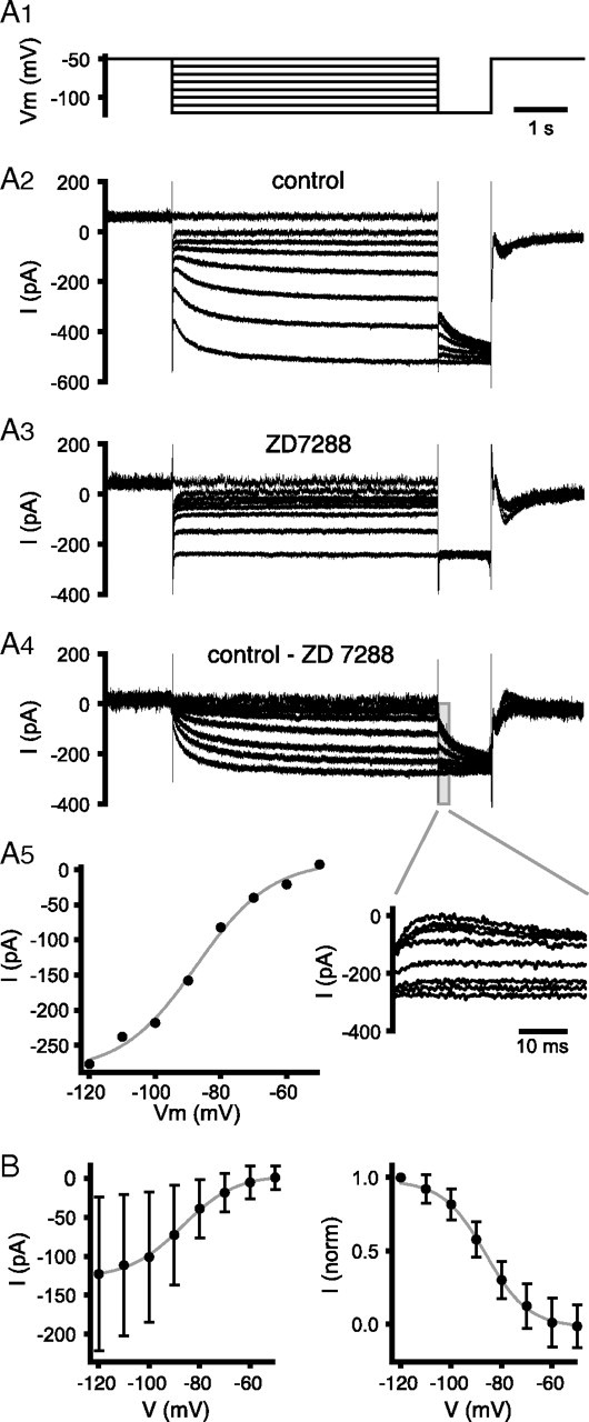
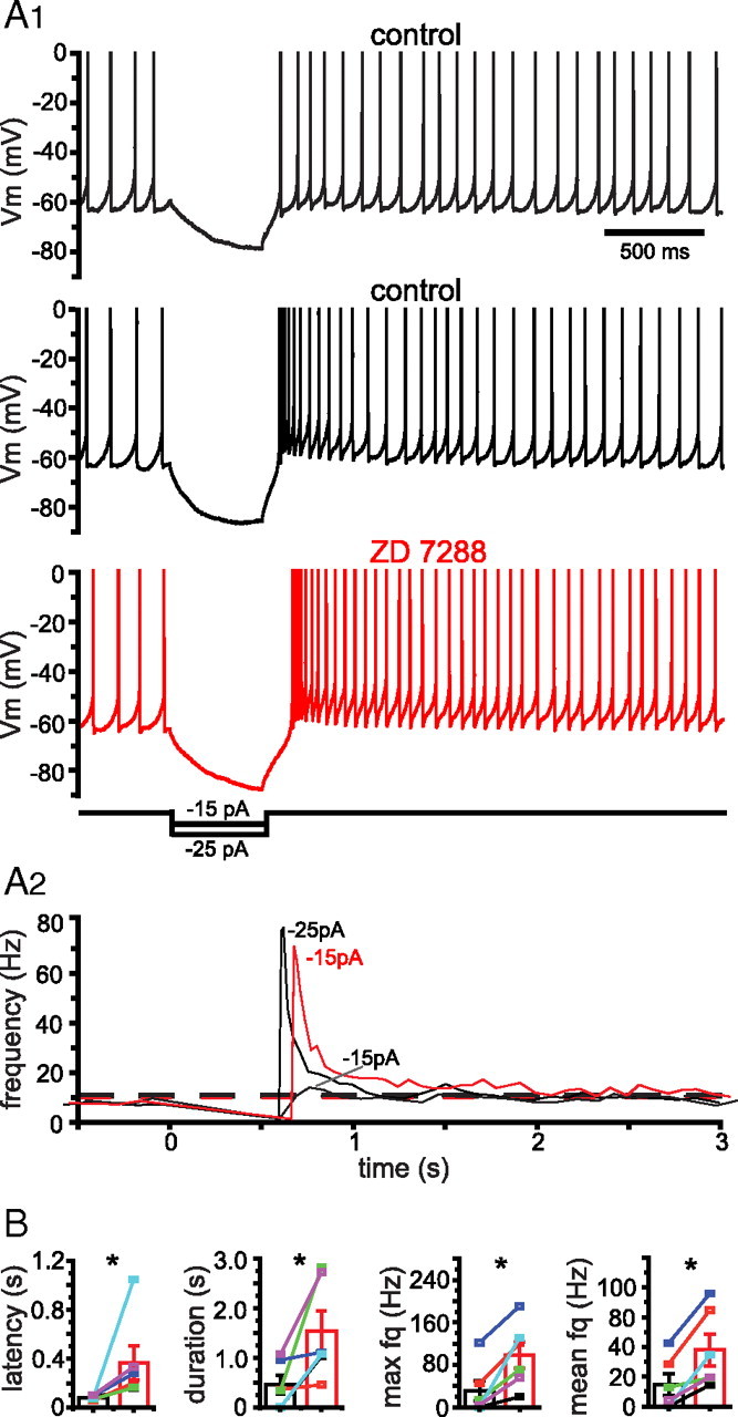
The activity patterns of subthalamic nucleus (STN) neurons are intimately linked to motor function and dysfunction and arise through the complex interaction of intrinsic properties and inhibitory and excitatory synaptic inputs. In many neurons, hyperpolarization-activated cyclic nucleotide-gated (HCN) channels play key roles in intrinsic excitability and synaptic integration both under normal conditions and in disease states. However, in STN neurons, which strongly express HCN channels, their roles remain relatively obscure. To address this deficit, complementary molecular and cellular electrophysiological, imaging, and computational approaches were applied to the rat STN. Molecular profiling demonstrated that individual STN neurons express mRNA encoding several HCN subunits, with HCN2 and 3 being the most abundant. Light and electron microscopic analysis showed that HCN2 subunits are strongly expressed and distributed throughout the somatodendritic plasma membrane. Voltage-, current-, and dynamic-clamp analysis, two-photon Ca(2+) imaging, and computational modeling revealed that HCN channels are activated by GABA(A) receptor-mediated inputs and thus limit synaptic hyperpolarization and deinactivation of low-voltage-activated Ca(2+) channels. Although HCN channels also limited the temporal summation of EPSPs, generated through two-photon uncaging of glutamate, this action was largely shunted by GABAergic inhibition that was necessary for HCN channel activation. Together the data demonstrate that HCN channels in STN neurons selectively counteract GABA(A) receptor-mediated inhibition arising from the globus pallidus and thus promote single-spike activity rather than rebound burst firing.


Figures

Figure 1.
Figure 1.
Expression of HCN channels. A, Gel from a single STN neuron that expressed HCN1, 2, and 3 mRNA. B, scRTPCR detection rate of HCN1–4 subunits in STN neurons (n = 16). C, Light micrograph of rat STN HCN2 immunoreactivity visualized with DAB and intensified by postfixation with osmium tetroxide. Arrowheads point to small immunoreactive elements in the neuropil. Neuronal somata (s) and small glial cells (g) were also weakly and strongly labeled, respectively. D, E, STN HCN2 immunoreactivity (as revealed by DAB without postfixation with osmium tetroxide) in a wild-type (WT) and a HCN2 knock-out (HCN2 KO) mouse. The pattern of labeling in the rat and WT mouse STN (labeled as for C) are similar. However, specific labeling was abolished in the STN of the HCN2 KO mouse.
Figure 2.
Figure 2.
HCN2 immunoreactivity is expressed across the somatodendritic axis of STN neurons. A, Electron micrograph of STN HCN2 immunoreactivity visualized with DAB. Reaction product (arrowheads) was observed in the soma (s) of a STN neuron and dendrites (d) of varying diameters. B, The density of HCN2 immunogold particles overlying the somatodendritic membrane (M) was similar across the somatodendritic axis and significantly higher than the densities overlying cytoplasmic (C) or nuclear compartments. This sample was taken from p41 tissue. C, D, Selective association of HCN2 immunogold particles (arrowheads) with the plasma membrane of small- (<1 μm) and large- (>1 μm) diameter dendritic (d; C) and somatic (s; D) compartments of STN neurons. In C, a terminal (*) with the morphology of a globus pallidus terminal forms symmetrical synaptic contacts (arrows) with a large-diameter immunoreactive dendrite. In these examples, immunoreactivity was absent from myelinated axons (a). *p < 0.05; box plot: box, 25%, 50%, and 75%; whiskers, 10–90%.
Figure 3.
Figure 3.
Effects of HCN channel blockade on excitability. A, Blockade of HCN channels with ZD7288 had no effect on autonomous activity or magnitude of action potential afterhyperpolarization (AHP) in a representative neuron (A1) or the sample population (A2, A3; individual neuron data represented by distinct colors). B, Blockade of HCN channels with ZD7288 did not affect the response of STN neurons to moderate depolarizing current injection either in the representative example neuron (B1) or across the sample population (B2). C, Blockade of HCN channels with ZD7288 (red) increased the degree of hyperpolarization produced by hyperpolarizing current injection in a representative neuron (C1) and across the sample population (C2, C3) compared to responses under control conditions (black). Effects on the degree of steady-state hyperpolarization measured at the end of 500 ms current injection were more consistent than effects on peak hyperpolarization. Action potentials are truncated at 0 mV; Vm, membrane potential; *p < 0.05.
Figure 4.
Figure 4.
Voltage dependence of HCN channel activation. A, Representative example. A1, Voltage-clamp waveform used to measure voltage dependence of HCN channel activation. A2–A4, Currents measured under control conditions (A2) and in the presence of ZD7288 (A3) and ZD7288-sensitive currents (A4) (inset shows tail currents evoked at −120 mV). A5, ZD7288-sensitive tail currents increased with progressive hyperpolarization from −60 mV. B, Absolute and normalized (norm) ZD7288-sensitive tail currents for the sample population. Vm, Membrane potential.
Figure 5.
Figure 5.
The impact of GABAA receptor-mediated inhibition is enhanced by blockade of HCN channels. A1, Response of a STN neuron under control conditions (black) and in the presence of ZD7288 (red) to an in vivo-like GABAA receptor-mediated synaptic conductance waveform (blue). A2, Zoom of epoch highlighted by blue rectangle in A1. Blockade of HCN channels increased inhibition of autonomously generated action potentials (control example 31 action potentials; ZD7288 example 25 action potentials) and the degree of hyperpolarization produced by the GABAA receptor-mediated synaptic conductance in both the representative example (A) and the sample population (B; individual cells denoted by distinct colors; sample mean and SD also indicated). Vm, Membrane potential; *p < 0.05.
Figure 6.
Figure 6.
HCN channels distributed across the somatodendritic axis most effectively counteract GABAergic inhibition. A, Somatic (black) and fourth (most distal) dendritic compartment (gray) responses of each STN neuron model to a GABAA receptor-mediated-synaptic conductance (top) applied to the fourth dendritic compartment. The number of action potentials generated during GABAergic inhibition was 41, 54, and 30 for the somatic, somatodendritic, and deficient models, respectively. B, Zoom of the epoch 2–2.4 s from the traces in A. During the period of inhibition the somatic and fourth dendritic compartments of the somatodendritic model (blue) were relatively depolarized compared to the somatic (green) and deficient (red) models. Whole-cell HCN and Nav channel currents were also greatest for the somatodendritic model. Vm, Membrane potential.
Figure 7.
Figure 7.
Blockade of HCN channels increases the intensity of rebound burst firing. A, Response of a STN neuron under control conditions (black) and in the presence of ZD7288 (red) to hyperpolarizing current injection. A1, From top to bottom, responses to −15 pA, −25 pA, and −15 pA, respectively. A2, Instantaneous frequency plots for each trace. Dashed line represents the mean instantaneous frequency + 3SD associated with autonomous activity. The degree of hyperpolarization and frequency of subsequent rebound activity in response to −15 pA were greatly enhanced by ZD7288. Application of −25 pA and −15 pA under control conditions and in the presence of ZD7288 produced similar degrees of hyperpolarization and rebound activity, respectively. B, Population data. Individual cells are represented by distinct colors. Population mean and SD are also indicated. Action potentials truncated at 0 mV; fq, frequency; Vm, membrane potential; *p < 0.05.
Figure 8.
Figure 8.
Blockade of HCN channels increases rebound activity of low-voltage-activated Ca2+ channels. A1, Left panel, 2PLSM image of a STN neuron with the site of a dendritic line scan indicated (green bar); right panel, voltage and Ca2+ indicator response to hyperpolarizing current injection under control conditions (black) and in the presence of ZD7288 (red). A2, A3, Population data. Individual cells denoted by distinct colors. Mean and SD are indicated. Rebound depolarization and rebound Ca2+ entry were enhanced by ZD7288. B1, Left panel, 2PLSM image of a STN neuron with the site of a dendritic line scan indicated (green bar); right panel, voltage and Ca2+ indicator response to GABAA receptor conductance injection under control conditions (black) and in the presence ZD7288 (red). The GABAA receptor conductance more effectively hyperpolarizes the membrane potential following blockade of HCN channels. B2, B3, Population data. Individual cells denoted by distinct colors. Mean and SD are indicated. Rebound depolarization and rebound Ca2+ entry were enhanced by application of ZD7288. Vm, Membrane potential; *p < 0.05.
Figure 9.
Figure 9.
HCN channels reduce the propensity for Cav3 channel-mediated rebound burst firing. A, Somatic responses of the somatic, somatodendritic, and deficient STN HCN models at the end and offset of somatic GABAA receptor synaptic conductance injection. The effectiveness of GABAA receptor-mediated hyperpolarization and subsequent rebound burst firing is enhanced by deficiency of HCN channels. B, Conductance (expressed as % g/gmax) of Cav3 channels for the three models at the end and offset of somatic GABAA receptor synaptic conductance injection. During inhibition the conductance of Cav3 channels is smallest in the HCN-deficient model. At the offset of inhibition the conductance of Cav3 channels is largest in the HCN-deficient model. Vm, Membrane potential.
Figure 10.
Figure 10.
Glutamatergic excitation is enhanced by blockade of HCN channels in a voltage-dependent manner. A1, 2PLSM image of a STN neuron together with sites of 2PLU of MNI-glutamate (1–3). A2, Somatic compound EPSPs generated at approximately −80 mV by 2PLU of MNI-glutamate (green) and associated Ca2+ dynamics at spine 1 and underlying dendritic shaft under control conditions (black) and in the presence of ZD7288 (red). The somatic EPSP was amplified by blockade of HCN channels, but associated Ca2+ dynamics were unaltered. B, Population data. Individual cells denoted by distinct colors. Mean and SD are indicated. The somatic EPSP integral generated at −80 mV and −70 mV by 2PLU of MNI-glutamate was enhanced and unaltered by application of ZD7288, respectively. Vm, Membrane potential; *p < 0.05.
Figure 11.
Figure 11.
GABAA receptor-mediated inhibition shunts the effect of HCN channels on excitatory synaptic integration. A1, 2PLSM image of a STN neuron together with sites of 2PLU of MNI-glutamate (green). A2, Somatic compound EPSPs generated by 2PLU of MNI-glutamate (green) during the injection of a 12 nS GABAA receptor-mediated conductance under control conditions (black) and in the presence of ZD7288 (red). In the presence of the GABAA conductance, blockade of HCN channels had little effect on the compound EPSP integral either in the example or the sample (A3; cells denoted by distinct colors; mean and SD indicated). B–D, Integration of three excitatory synaptic conductances (AMPA, 1 nS; NMDA, 5 nS) delivered to the distal dendritic compartment at 50 ms intervals was analyzed for each STN model under three regimes of inhibition. B, Somatic hyperpolarizing current was injected to generate to steady-state somatic hyperpolarization of −80 mV in each model (somatic, −57.9 pA; somatodendritic, −49.1 pA; deficient, −20.2 pA). C, Somatic GABAergic conductances (somatic, 14.5 nS; somatodendritic, 12.3 nS; deficient, 5.05 nS) were injected to generate steady-state somatic hyperpolarization of −80 mV. D, An identical somatic GABAergic conductance (12.3 nS) was injected for each model. When the GABAergic conductance was identical EPSP integration was not affected by the presence or absence of HCN channels although Vm was more hyperpolarized in the deficient model. Vm, Membrane potential.

References

    1. Afsharpour S. Light microscopic analysis of Golgi-impregnated rat subthalamic neurons. J Comp Neurol. 1985;236:1–13. - PubMed
    1. Angelo K, London M, Christensen SR, Häusser M. Local and global effects of Ih distribution in dendrites of mammalian neurons. J Neurosci. 2007;27:8643–8653. - PMC - PubMed
    1. Atherton JF, Wokosin DL, Ramanathan S, Bevan MD. Autonomous initiation and propagation of action potentials in neurons of the subthalamic nucleus. J Physiol. 2008;586:5679–5700. - PMC - PubMed
    1. Baruscotti M, DiFrancesco D. Pacemaker channels. Ann N Y Acad Sci. 2004;1015:111–121. - PubMed
    1. Baufreton J, Bevan MD. D2-like dopamine receptor-mediated modulation of activity-dependent plasticity at GABAergic synapses in the subthalamic nucleus. J Physiol. 2008;586:2121–2142. - PMC - PubMed

Publication types

MeSH terms

LinkOut - more resources