. 2011 Jan 1;186(1):516-26.
doi: 10.4049/jimmunol.1000955. Epub 2010 Nov 24.

Chemoattractant-induced signaling via the Ras-ERK and PI3K-Akt networks, along with leukotriene C4 release, is dependent on the tyrosine kinase Lyn in IL-5- and IL-3-primed human blood eosinophils

Affiliations

Chemoattractant-induced signaling via the Ras-ERK and PI3K-Akt networks, along with leukotriene C4 release, is dependent on the tyrosine kinase Lyn in IL-5- and IL-3-primed human blood eosinophils

Yiming Zhu et al. J Immunol. .

Abstract

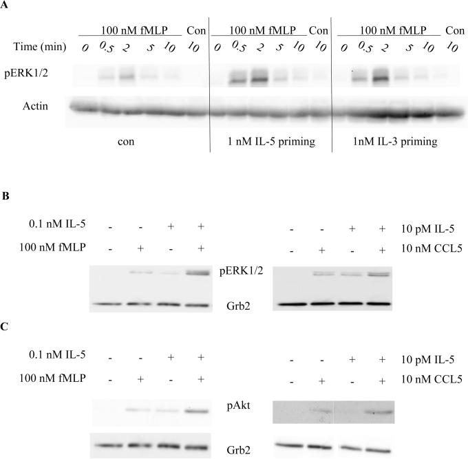
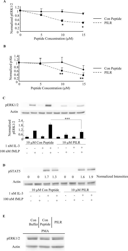
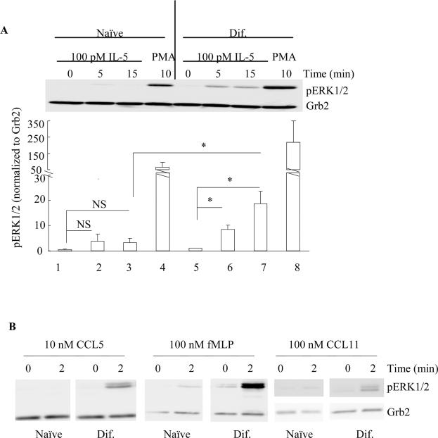
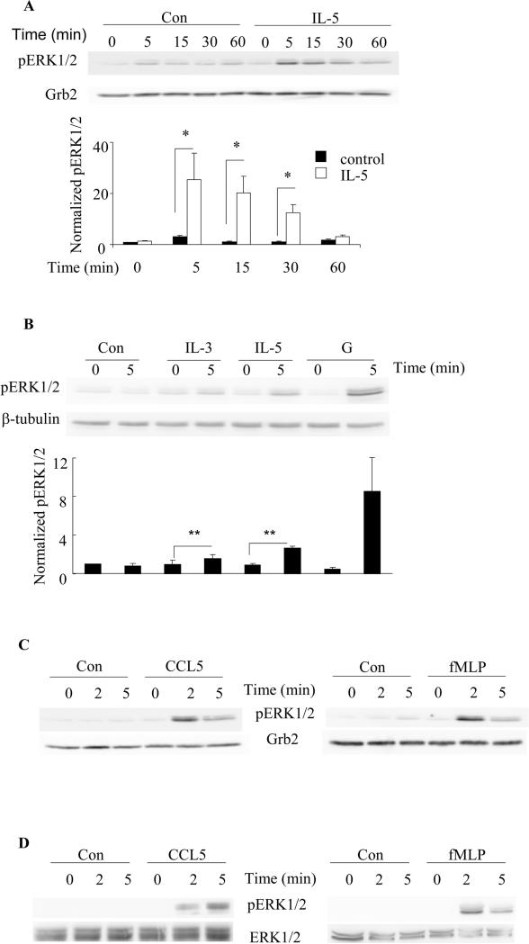
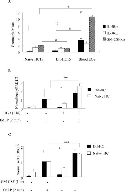
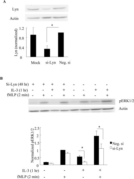
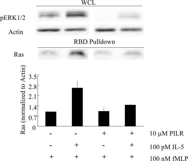
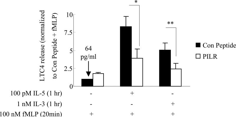
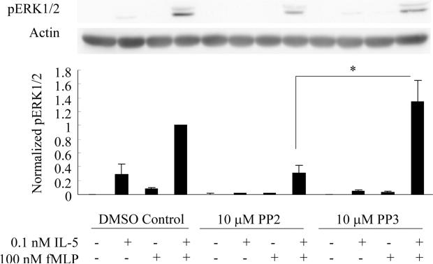
Human blood eosinophils exhibit a hyperactive phenotype in response to chemotactic factors after cell "priming" with IL-5 family cytokines. Earlier work has identified ERK1/2 as molecular markers for IL-5 priming, and in this article, we show that IL-3, a member of the IL-5 family, also augments fMLP-stimulated ERK1/2 phosphorylation in primary eosinophils. Besides ERK1/2, we also observed an enhancement of chemotactic factor-induced Akt phosphorylation after IL-5 priming of human blood eosinophils. Administration of a peptide antagonist that targets the Src family member Lyn before cytokine (IL-5/IL-3) priming of blood eosinophils inhibited the synergistic increase of fMLP-induced activation of Ras, ERK1/2 and Akt, as well as the release of the proinflammatory factor leukotriene C(4). In this study, we also examined a human eosinophil-like cell line HL-60 clone-15 and observed that these cells exhibited significant surface expression of IL-3Rs and GM-CSFRs, as well as ERK1/2 phosphorylation in response to the addition of IL-5 family cytokines or the chemotactic factors fMLP, CCL5, and CCL11. Consistent with the surface profile of IL-5 family receptors, HL-60 clone-15 recapitulated the enhanced fMLP-induced ERK1/2 phosphorylation observed in primary blood eosinophils after priming with IL-3/GM-CSF, and small interfering RNA-mediated knockdown of Lyn expression completely abolished the synergistic effects of IL-3 priming on fMLP-induced ERK1/2 phosphorylation. Altogether, our data demonstrate a central role for Lyn in the mechanisms of IL-5 family priming and suggest that Lyn contributes to the upregulation of the Ras-ERK1/2 and PI3K-Akt cascades, as well as the increased leukotriene C(4) release observed in response to fMLP in "primed" eosinophils.


Figures

Figure 1
Figure 1. IL-5 and IL-3 prime chemotactic factor-induced phosphorylation of ERK1/2 and Akt
A. Purified human blood eosinophils were primed with control buffer, IL-5 or IL-3 for 1 hr, followed by stimulation with fMLP or control for the indicated time points. Cell lysates were processed by SDS-PAGE and immunoblotted for pERK1/2 and actin (A, N=3) as detailed under Materials and Methods. B and C. Eosinophils were primed with control buffer or IL-5 for 1 hr followed by a two-min stimulation with fMLP or CCL5. Cell lysates were subjected to SDS-PAGE and immunoblotted for pERK1/2 (B, N=5), pAkt (C, N=4) and Grb2.
Figure 2
Figure 2. PILR inhibits fMLP-stimulated ERK1/2 and Akt phosphorylation in human blood eosinophils primed with IL-5 family members
Blood eosinophils were preincubated with the Lyn recruitment antagonist PILR or control peptide for 30 min prior to priming with 100 pM IL-5 (A, B. D) or 1 nM IL-3(C) for 1 hr, followed by stimulation with 100 nM fMLP for 2 min. Phosphorylation of ERK1/2, Akt and STAT5 was assessed by SDS-PAGE and immunoblotting as detailed under Materials and Methods. Levels of actin were used as loading controls in all blots. Band intensities of pERK1/2 (A, *p<0.002, N=6, PILR vs. Con Peptide) and pAkt (B, **p<0.05, N=4, PILR vs. Con Peptide) were quantified by densitometry analysis and normalized to that of actin on the same blot. Data are summarized as the mean ± (SEM). C. Representative immunoblots are shown for five independent experiments. Data are summarized in the lower panel as the mean ± (SEM). ***p<0.05, N=5. D. Immunoblots of pSTAT5 and actin are shown as representatives of three independent experiments. Normalized band intensities expressed as fold induction are shown under the pSTAT5 immunoblot. E. Eosinophils were pretreated with control buffer, 10 μM control peptide or PILR for 30 min prior to the stimulation with 10 ng/ml PMA for 10 min. Representative immunoblots of pERK1/2 and actin of three independent experiments are shown.
Figure 3
Figure 3. Differential responses of ERK1/2 phosphorylation in the naïve and dif-HC15 in response to IL-5 and chemotactic factors fMLP and CCL5
A. The naïve (lane 1–4) and dif-HC15 (lane 5–8) cells were stimulated with 100 pM IL-5 for 0 min (lane 1, 5), 5 min (lane 2, 6), 15 min (lane 3, 7) or 10 ng/ml PMA for 10 min (lane 4, 8). Band intensities of pERK1/2 were normalized to that of Grb2, and then normalized to that of lane 1. Data are summarized as the mean ± (SEM) from three independent experiments. NS=Not Significant. * p<0.05, N=3. B. Naïve and dif-HC15 cells were treated with 10 nM CCL5, 100 nM fMLP or 100 nM CCL11 for 0 or 2 min. Cell extracts were then prepared and immunoblotted as detailed under Materials and Methods. Representative immunoblots of pERK1/2 and Grb2 from three independent experiments are shown.
Figure 4
Figure 4. Kinetics of ERK1/2 phosphorylation in the dif-HC15 cells in response to IL-5 family cytokines and chemotactic factors fMLP and CCL5
A. The dif-HC15 were stimulated with 100 pM IL-5 or control vehicle for the indicated times. Cell lysates were subject to SDS-PAGE and immunoblotting for pERK1/2 and Grb2. The upper panel shows representative immunoblots of pERK1/2 and Grb2 of six independent experiments. ERK1/2 phosphorylation levels were normalized to that of Grb2, and IL-5-treated at time 0 was set to be 1. Data are summarized as the mean ± (SEM) of six experiments in the lower panel. *p<0.05, N=6. A two-way ANOVA was used for the statistical analysis. B. The dif-HC15 cells were stimulated with control (Con), 1nM IL-3, IL-5 or GM-CSF (G) for 0 or 5 min. The upper panel shows representative immunoblots of pERK1/2 and β-tubulin of three independent experiments. The lower panel is the summary of the mean ± (SEM). The control-treated cells at time 0 was set to be 1. **p<0.05 N=3. The dif-HC15 cells (C) and human blood eosinophils (D) were stimulated with 10 nM CCL5 or 100 nM fMLP for 0, 2 or 5 min. Representative immunoblots of pERK1/2 and the corresponding loading controls of three independent experiments are shown.
Figure 5
Figure 5. Surface expression profile of IL-5 family receptors and priming in HC15 cells
A. Naïve HC15, dif-HC15 and human blood eosinophils (Blood EOS) were examined for surface expression of IL-5Rα, IL-3Rα and GM-CSFRα using flow cytometry as described under Materials and Methods. Geometric means of the α subunits were normalized by subtracting the geometric means of the corresponding IgG control and are presented as the mean ± (SEM); similar results were obtained when the data were normalized to a positive control (primary eosinophils) The data are summarized from four independent experiments for each receptor. #p<0.006, N=4. HC15 cells were primed with 1 nM of IL-3 (B) or GM-CSF (C) for 1 hr, and stimulated with 100 nM fMLP for 2 min. Protein levels of pERK1/2 and actin were detected by immunoblotting. Band intensities of pERK1/2 were normalized to that of actin. Cells treated with fMLP alone were set to be 1 in both panels. The data are presented as the mean ± (SEM). *p<0.02 **p<0.003 N=6, ***p<0.004 N=3.
Figure 6
Figure 6. Suppressing Lyn expression using siRNA inhibits the enhanced ERK1/2 phosphorylation in response to fMLP following IL-3 priming in the naïve HC15
Naïve HC15 cells were transfected with no siRNA (Mock), siRNA against Lyn (si-Lyn) or negative control siRNA (Neg. si) for 48 hr as detailed under Materials and Methods. The expression levels of Lyn were examined by immunoblotting (A, upper panel) and summarized as the mean ± (SEM) from four independent experiments (A, lower panel). Following Lyn knockdown for 48 hr, the cells were treated with 1 nM IL-3 for 1 hr and then stimulated with 100 nM fMLP for 2 min. Representative immunoblots of pERK1/2 and actin (B, upper panel), and the summary of the mean ± (SEM) from four independent experiments (B, lower panel) are shown. *p<0.02, N=4.
Figure 7
Figure 7. PILR inhibits fMLP-stimulated Ras activation following IL-5 priming in human blood eosinophils
Human primary eosinophils were preincubated with 10 μM control peptide or PILR for 30 min. The cells were then primed with 100 pM IL-5 or buffer control for 1 hr followed by 100 nM fMLP for 2 min. Cells lysates were subjected to the RBD pull-down assay as detailed under Materials and Methods. An aliquot of the whole cell lysate (WCL) from each treatment was removed to assess levels of pERK1/2 and actin (upper panel). The levels of active Ras were detected in the pull-down samples via immunobloting (upper panel) and normalized to that of actin in the whole cell lysates. The mean ± (SEM) from four independent experiments are summarized in the lower panel.
Figure 8
Figure 8. PILR inhibits LTC4 release in response to fMLP stimulation in IL-5/IL-3-primed human blood eosinophils
Human blood eosinophils were preincubated with 10 μM control peptide or PILR for 30 min. Cells were then primed with control buffer, 100 pM IL-5, or 1 nM IL-3 for 1 hr followed by a 20 min stimulation with 100 nM fMLP. Supernatants were then collected and assayed for immunoreactive LTC4 as described under Materials and Methods. The data are normalized to the LTC4 released from the leftmost bar and presented as fold increases. The dark bars and open bars represent the pretreatment with control peptide and PILR, respectively. Seven independent experiments are summarized and presented as the mean ± (SEM). * p<0.04 **p<0.02, N=7.
Figure 9
Figure 9. Src inhibitor PP2 attenuates fMLP-induced ERK1/2 phosphorylation in IL-5-primed human blood eosinophils
Human blood eosinophils were pretreated with 10 μM PP2, PP3 or vehicle (DMSO) control for 30 min. Subsequent to this treatment, the cells were primed with 100 pM IL-5 for 1 hr followed by the addition of 100 nM fMLP for 2 min. Cell lysates were collected and subjected to SDS-PAGE. The phosphorylation status of ERK1/2 (pERK1/2) was detected by immunoblotting and actin was used as a loading control. Representative immunoblots of pERK1/2 and actin are shown in the upper panel. Fold increases in pERK1/2 were ascertained by normalizing the band intensity of pERK1/2 to the corresponding actin control. Six independent experiments are summarized as the mean ± (SEM) in the lower panel. *p<0.04, N=6.

References

    1. Moore WC, Peters SP. Severe asthma: an overview. J Allergy Clin Immunol. 2006;117 - PubMed
    1. Sims JM. An overview of asthma. Dimens Crit Care Nurs. 2006;25 - PubMed
    1. Balkissoon R. Asthma overview. Prim Care. 2008;35 - PubMed
    1. Arm JP, Lee TH. The pathobiology of bronchial asthma. Adv Immunol. 1992;51 - PubMed
    1. Frigas E, Gleich GJ. The eosinophil and the pathophysiology of asthma. J Allergy Clin Immunol. 1986;77 - PubMed

Publication types

MeSH terms