

Follistatin regulates germ cell nest breakdown and primordial follicle formation
- PMID: 21106872
- PMCID: PMC3037165
- DOI: 10.1210/en.2010-0950
Follistatin regulates germ cell nest breakdown and primordial follicle formation
Abstract
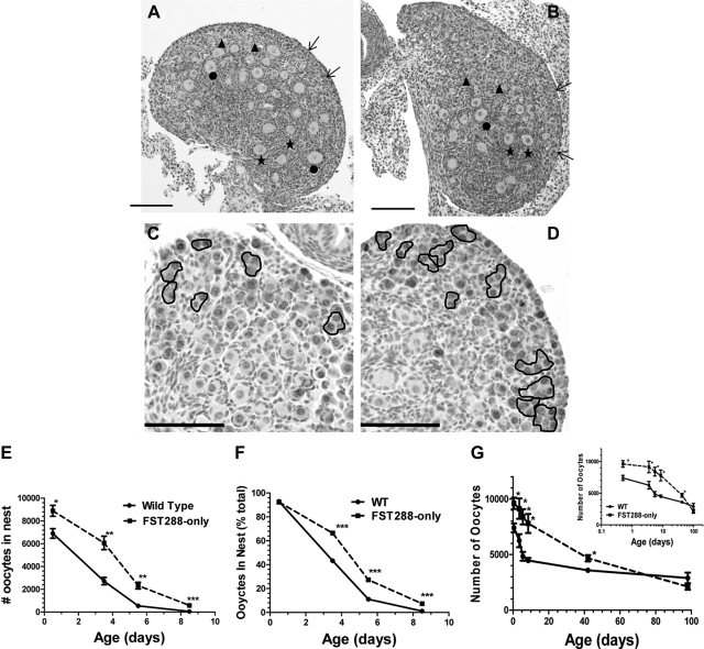
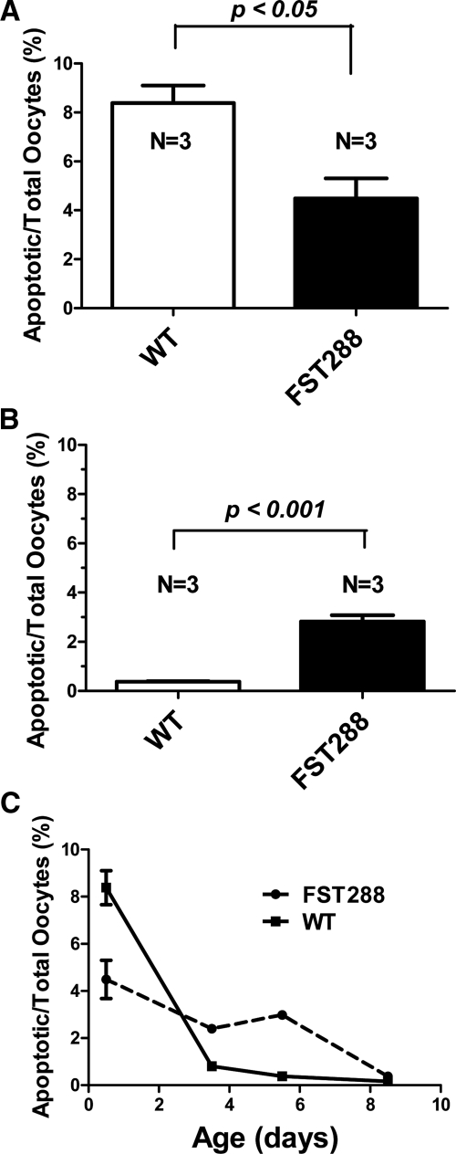
Follistatin (FST) is an antagonist of activin and related TGFβ superfamily members that has important reproductive actions as well as critical regulatory functions in other tissues and systems. FST is produced as three protein isoforms that differ in their biochemical properties and in their localization within the body. We created FST288-only mice that only express the short FST288 isoform and previously reported that females are subfertile, but have an excess of primordial follicles on postnatal day (PND) 8.5 that undergo accelerated demise in adults. We have now examined germ cell nest breakdown and primordial follicle formation in the critical PND 0.5-8.5 period to test the hypothesis that the excess primordial follicles derive from increased proliferation and decreased apoptosis during germ cell nest breakdown. Using double immunofluorescence microscopy we found that there is virtually no germ cell proliferation after birth in wild-type or FST288-only females. However, the entire process of germ cell nest breakdown was extended in time (through at least PND 8.5) and apoptosis was significantly reduced in FST288-only females. In addition, FST288-only females are born with more germ cells within the nests. Thus, the excess primordial follicles in FST288-only mice derive from a greater number of germ cells at birth as well as a reduced rate of apoptosis during nest breakdown. These results also demonstrate that FST is critical for normal regulation of germ cell nest breakdown and that loss of the FST303 and/or FST315 isoforms leads to excess primordial follicles with accelerated demise, resulting in premature cessation of ovarian function.
Figures
Similar articles
-
Follistatin288 Regulates Germ Cell Cyst Breakdown and Primordial Follicle Assembly in the Mouse Ovary.PLoS One. 2015 Jun 15;10(6):e0129643. doi: 10.1371/journal.pone.0129643. eCollection 2015. PLoS One. 2015. PMID: 26076381 Free PMC article.
-
The follistatin-288 isoform alone is sufficient for survival but not for normal fertility in mice.Endocrinology. 2010 Mar;151(3):1310-9. doi: 10.1210/en.2009-1176. Epub 2009 Dec 23. Endocrinology. 2010. PMID: 20032047 Free PMC article.
-
Germ cell dynamics during nest breakdown and formation of the primordial follicle pool in the domestic turkey (Meleagris gallopavo).Poult Sci. 2020 May;99(5):2746-2756. doi: 10.1016/j.psj.2019.12.050. Epub 2020 Mar 20. Poult Sci. 2020. PMID: 32359612 Free PMC article.
-
From primordial germ cells to primordial follicles: a review and visual representation of early ovarian development in mice.J Ovarian Res. 2016 Jun 21;9(1):36. doi: 10.1186/s13048-016-0246-7. J Ovarian Res. 2016. PMID: 27329176 Free PMC article. Review.
-
The primordial pool of follicles and nest breakdown in mammalian ovaries.Mol Hum Reprod. 2009 Dec;15(12):795-803. doi: 10.1093/molehr/gap073. Epub 2009 Aug 26. Mol Hum Reprod. 2009. PMID: 19710243 Free PMC article. Review.
Cited by
-
Conservation of oocyte development in germline cysts from Drosophila to mouse.Elife. 2022 Nov 29;11:e83230. doi: 10.7554/eLife.83230. Elife. 2022. PMID: 36445738 Free PMC article.
-
Blocking estrogen-induced AMH expression is crucial for normal follicle formation.Development. 2021 Mar 19;148(6):dev197459. doi: 10.1242/dev.197459. Development. 2021. PMID: 33658225 Free PMC article.
-
Follistatin288 Regulates Germ Cell Cyst Breakdown and Primordial Follicle Assembly in the Mouse Ovary.PLoS One. 2015 Jun 15;10(6):e0129643. doi: 10.1371/journal.pone.0129643. eCollection 2015. PLoS One. 2015. PMID: 26076381 Free PMC article.
-
Simultaneous gene deletion of gata4 and gata6 leads to early disruption of follicular development and germ cell loss in the murine ovary.Biol Reprod. 2014 Jul;91(1):24. doi: 10.1095/biolreprod.113.117002. Epub 2014 Jun 4. Biol Reprod. 2014. PMID: 24899573 Free PMC article.
-
Regulation of the ovarian reserve by members of the transforming growth factor beta family.Mol Reprod Dev. 2012 Oct;79(10):666-79. doi: 10.1002/mrd.22076. Epub 2012 Sep 11. Mol Reprod Dev. 2012. PMID: 22847922 Free PMC article. Review.
References
-
- Tortoriello DV, Sidis Y, Holtzman DA, Holmes WE, Schneyer AL. 2001. Human follistatin-related protein: a structural homologue of follistatin with nuclear localization. Endocrinology 142:3426–3434 - PubMed
-
- Sugino K, Kurosawa N, Nakamura T, Takio K, Shimasaki S, Ling N, Titani K, Sugino H. 1993. Molecular heterogeneity of follistatin, an activin-binding protein. Higher affinity of the carboxyl-terminal truncated forms for heparan sulfate proteoglycans on the ovarian granulosa cell. J Biol Chem 68:15579–15587 - PubMed
-
- Vale W, Rivier C, Hsueh A, Campen C, Meunier H, Bicsak T, Vaughan J, Corrigan A, Bardin W, Sawchenko P, Spiess J, Rivier J. 1988. Chemical and biological characterization of the inhibin family of protein hormones. Rec Prog Horm Res 44:1–34 - PubMed
-
- Mather JP, Moore A, Li RH. 1997. Activins, inhibins, and follistatins: further thoughts on a growing family of regulators. Proc Soc Exp Biol Med 215:209–222 - PubMed
Publication types
MeSH terms
Substances
Grants and funding
LinkOut - more resources
Full Text Sources
Molecular Biology Databases
Miscellaneous
