. 2011 Jan;152(1):138-50.
doi: 10.1210/en.2010-0791. Epub 2010 Nov 24.

Glucocorticoids differentially regulate the expression of CRFR1 and CRFR2α in MIN6 insulinoma cells and rodent islets

Affiliations

Glucocorticoids differentially regulate the expression of CRFR1 and CRFR2α in MIN6 insulinoma cells and rodent islets

M O Huising et al. Endocrinology. 2011 Jan.

Abstract

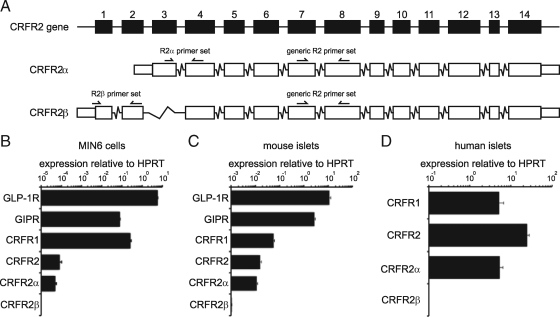
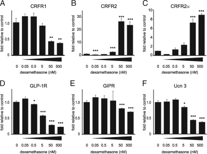
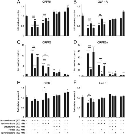
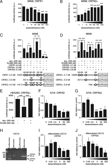
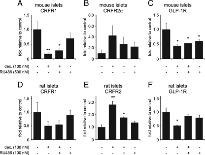
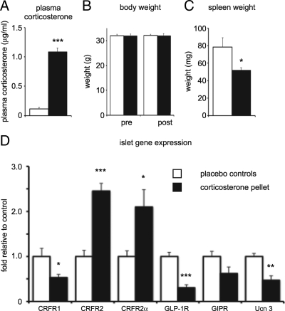
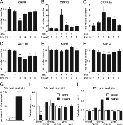
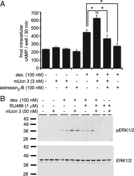
Urocortin 3 (Ucn 3), member of the corticotropin-releasing factor (CRF) family of peptide hormones, is released from β-cells to potentiate insulin secretion. Ucn 3 activates the CRF type-2 receptor (CRFR2) but does not activate the type-1 receptor (CRFR1), which was recently demonstrated on β-cells. While the direct actions of Ucn 3 on insulin secretion suggest the presence of cognate receptors within the islet microenvironment, this has not been established. Here we demonstrate that CRFR2α is expressed by MIN6 insulinoma cells and by primary mouse and human islets, with no detectable expression of CRFR2β. Furthermore, stimulation of MIN6 cells or primary mouse islets in vitro or in vivo with glucocorticoids (GCs) robustly and dose-dependently increases the expression of CRFR2α, while simultaneously inhibiting the expression of CRFR1 and incretin receptors. Luciferase reporters driven by the mouse CRFR1 or CRFR2α promoter in MIN6 cells confirm these differential effects of GCs. In contrast, GCs inhibit CRFR2α promoter activity in HEK293 cells and inhibit the expression of CRFR2β in A7r5 rat aortic smooth muscle cells and differentiated C2C12 myotubes. These findings suggest that the GC-mediated increase of CRFR2α depends on the cellular context of the islet and deviates from the GC-mediated suppression of CRFR1 and incretin receptors. Furthermore, GC-induced increases in CRFR2α expression coincide with increased Ucn 3-dependent activation of cAMP and MAPK pathways. We postulate that differential effect of GCs on the expression of CRFR1 and CRFR2α in the endocrine pancreas represent a mechanism to shift sensitivity from CRFR1 to CRFR2 ligands.


Figures

Figure 1
Figure 1
The α isoform of CRFR2 is expressed in the MIN6 insulinoma cell line and in primary mouse and human islets. The use of primers designed to discriminate between mouse CRFR2α and CRFRβ isoforms (A) reveals that CRFR2α is the major CRFR2 isoform expressed in MIN6 cells (B). MIN6 cells express CRFR2 at relatively low abundance compared with the expression of incretin receptors and CRFR1. Primary mouse islets also express the CRFR2α isoform at levels that approach CRFR1 in abundance (C). A similar strategy for the design of isoform-specific primers revealed that CRFR2α is the major isoform of CRFR2 that is expressed in human islets as well (D). Boxes in A represent exons and are drawn to scale. Data are expressed relative to HPRT. Error bars in B and C indicate the SE of quadruplicate treatments, error bars in D reflect the SE across 20 individual donors.
Figure 2
Figure 2
GCs dose-dependently inhibit CRFR1 and increase CRFR2 expression in MIN6 cells. Stimulation of MIN6 cells for 12 h with the synthetic GC dexamethasone dose-dependently inhibits the expression of CRFR1 (A). In contrast, CRFR2α expression is robustly and dose-dependently increased by dexamethasone, as measured with generic (B) or CRFR2α-selective (C) qRT-PCR primers. The expression of GLP-1R (D), GIPR (E), and Ucn 3 (F) are inhibited by dexamethasone in a pattern similar to CRFR1. Data are normalized to HPRT expression and expressed relative to untreated controls. Error bars indicate the SE of quadruplicate treatments, asterisks indicate statistically significant differences with controls (*, P < 0.05; **, P < 0.01; ***, P < 0.001).
Figure 3
Figure 3
The GC-mediated transcriptional changes in MIN6 cells are GR-mediated. MIN6 cells were stimulated for 12 h with the GR-selective agonist dexamethasone, the MR-selective agonist aldosterone, or the general GR/MR agonist hydrocortisone alone or in combination with RU486 (GR antagonist) or spironolactone (MR antagonist) as indicated. Dexamethasone and hydrocortisone robustly inhibit the expression of CRFR1 (A) and GLP-1R (B), which is negated by co-administration of RU486, but not spironolactone, both at equimolar levels. Expression of neither gene is affected by aldosterone. In contrast, the expression of CRFR2, as detected by generic (C) or CRFR2α-specific (D) primers, is potently increased by dexamethasone and hydrocortisone. These increases are partially prevented by RU486, but not spironolactone, indicating that these changes are GR-dependent. The expression of GIPR (E) and Ucn 3 (F) is inhibited in a pattern that reminisces the expression of CRFR1 and GLP-1R, although this inhibition is less robust. Data are normalized to HPRT expression and expressed relative to untreated controls. Error bars indicate the SE of quadruplicate treatments, asterisks indicate statistically significant differences with controls unless indicated otherwise (*, P < 0.05; **, P < 0.01; ***, P < 0.001).
Figure 4
Figure 4
CRFR1- and CRFR2α promoter-driven luciferase activity is regulated oppositely in MIN6 cells and GC-mediated expression of CRFR2 is dependent on cellular context. MIN6 cells transfected with a luciferase reporter construct under control of the putative CRFR1 (A) or CRFR2α (B) promoters display opposite, dose-dependent effects on luciferase activity after 12-hour stimulation with increasing doses of dexamethasone. The opposite effects of dexamethasone on CRFR1 and CRFR2α promoter activity are fully inhibited by the GR-selective antagonist RU486 (C). CRFR2β promoter activity in MIN6 cells is relatively low, and is inhibited by dexamethasone (C). MIN6 cells transfected with one of three successively shorter fragments of the putative CRFR2α promoter consistently demonstrate dexamethasone-dependent increases in luciferase activity that are fully inhibited by co-administration of the GR-selective antagonist RU486 (D). Predicted GREs consisting of two palindromic half sites are represented by open circles. By contrast, CRFR2α promoter-driven luciferase activity in HEK293 cells is inhibited dose-dependently by dexamethasone, which is prevented by co-administration of RU486 (E). Stimulation of rat A7r5 aortic smooth muscle cells with dexamethasone for 12 h dose-dependently inhibits the expression of CRFR2β, as measured with generic CRFR2 (F) and CRFR2β-specific (G) primers. CRFR2α was not detected in A7r5 cells. Undifferentiated mouse C2C12 cells express low levels of CRFR2α (91 bp) but switch to express CRFR2β (294 bp) when differentiated (H). The approximately 300-bp species that appears in some CRFR2α lanes is an artifact resulting from the mis-priming of the mouse CRFR2α primers to laminin 5a cDNA, and has so far only been observed in C2C12 cells. Expression of CRFR2β in differentiated C2C12 myotubes is inhibited dose-dependently after stimulation for 12 h with increasing doses of dexamethasone, as measured with generic CRFR2 (I) and CRFR2β-specific (J) primers. Data in F, G, I, and J are normalized to HPRT expression and expressed relative to untreated controls. Error bars indicate the SE of triplicate (A–G) or quadruplicate (I and J) treatments, asterisks indicate statistically significant differences with controls unless indicated otherwise (*, P < 0.05; **, P < 0.01; ***, P < 0.001).
Figure 5
Figure 5
GCs differentially regulate the expression of CRFR2 and CRFR1 and GLP-1R in primary mouse and rat islets. Primary mouse (A–C) or rat (D–F) islets were stimulated for 12 h with dexamethasone and/or the GR-selective antagonist RU486 as indicated. Dexamethasone inhibited the expression of CRFR1 in mouse islets (A) and tended to inhibit CRFR1 expression in rat islets (D). In contrast, CRFR2 expression was increased in both mouse (B) and rat (E) islets, although the GC-induced increase of CRFR2α in mouse islets narrowly failed to reach statistical significance (one-sided P = 0.051). The expression of GLP-1R is inhibited by dexamethasone in both mouse (C) and rat (F) islets, although RU486 fails to fully revert the GC-mediated suppression of mouse GLP-1R and even slightly inhibits the expression of GLP-1R itself for reasons not entirely understood. Data are normalized to HPRT expression and expressed relative to untreated controls. Error bars indicate the SE of quadruplicate (A–C) or triplicate (D–F) treatments, asterisks indicate statistically significant differences with controls (*, P < 0.05; **, P < 0.01).
Figure 6
Figure 6
GCs differentially affect islet CRFR1 and CRFR2 expression in vivo. Corticosterone pellets were implanted subcutaneously in wild-type mice, leading to significantly elevated plasma corticosterone levels (A). Total body weight was not affected by pellet implantation (B), but spleen weight was significantly reduced (C). The islet gene expression profile reveals GC-induced inhibitions of CRFR1, GLP-1R, GIPR, and Ucn 3 expression, while the expression of CRFR2α is increased (D). Gene expression data are normalized to HPRT expression and expressed relative to placebo controls. Error bars indicate the SE of five (placebo group) or six (corticosterone group) animals, asterisks indicate statistically significant differences with placebo controls (*, P < 0.05; **, P < 0.01; ***, P < 0.001).
Figure 7
Figure 7
Acute GC exposure changes CRFR1 and CRFR2 expression in MIN6 cells and in islets in vivo. MIN6 cells were exposed to 100 nm dexamethasone, which was removed by triple washing after 1 h. This short exposure to dexamethasone suffices to inhibit CRFR1 expression (A) and increase CRFR2 expression (B and C). Expression of GLP-1R (D) and Ucn 3 (F) but not GIPR (E) is inhibited by dexamethasone as well. All GC-mediated effects on gene expression are transient, reach maximum amplitude between 3 h and 6 h, and normalize 9 h after the initiation of dexamethasone stimulation. To assess whether a similar acute and transient GC exposure would elicit gene expression changes in islets in vivo, we assessed islet gene expression after 30 min of restraint stress. Three hours after restraint, plasma cortisol levels are still significantly elevated in stressed animals compared with controls (G), and the islet expression of CRFR1 and GLP-1R is significantly reduced (H). However, 12 h after acute restraint, no significant changes in islet gene expression remain (I). Error bars on MIN6 data (A–F) indicate the SE of quadruplicates, whereas error bars reflect the SE of six controls and seven restrained animals (G and H) or five controls and four restrained animals (I). Asterisks indicate statistically significant differences with placebo controls (*, P < 0.05; **, P < 0.01; ***, P < 0.001).
Figure 8
Figure 8
GC-mediated up-regulation of CRFR2 increases responsiveness to Ucn 3. The relatively low expression of CRFR2α in MIN6 cells is insufficient to induce the accumulation of intracellular cAMP in response to mUcn 3 (A). However, 12-h pretreatment with 100 nm dexamethasone instills MIN6 cells with the ability to respond to mUcn 3 with increased levels of intracellular cAMP. Note that basal levels of cAMP are elevated after dexamethasone pre-treatment. This may be caused in part by endogenous Ucn 3 expressed in, and secreted from MIN6 cells, as stimulation with 100 nm of the CRFR2-selective antagonist astressin2-B inhibits cAMP levels to well below the initial baseline, suggesting the presence of Ucn 3 tone. Similarly, MIN6 cells do not respond to a short (5 min) stimulation with 50 nm mUcn 3 with phosphorylation of ERK1/2, unless pretreated overnight with 100 nm dexamethasone (B). Co-administration of the GR antagonist RU486 during dexamethasone pre-treatment interferes with the ability of MIN6 cells to gain responsivity to Ucn 3. Total ERK1/2 levels were equal among all wells. Error bars in A indicate the SE of triplicate treatments, asterisks indicate statistically significant differences compared with controls that received dexamethasone (*, P < 0.05).

Similar articles

Cited by

References

    1. Strowski MZ, Parmar RM, Blake AD, Schaeffer JM 2000 Somatostatin inhibits insulin and glucagon secretion via two receptors subtypes: an in vitro study of pancreatic islets from somatostatin receptor 2 knockout mice. Endocrinology 141:111–117 - PubMed
    1. Baggio LL, Drucker DJ 2007 Biology of incretins: GLP-1 and GIP. Gastroenterology 132:2131–2157 - PubMed
    1. Hansotia T, Drucker DJ 2005 GIP and GLP-1 as incretin hormones: lessons from single and double incretin receptor knockout mice. Regul Pept 128:125–134 - PubMed
    1. Gilon P, Henquin JC 2001 Mechanisms and physiological significance of the cholinergic control of pancreatic beta-cell function. Endocr Rev 22:565–604 - PubMed
    1. Huising MO, van der Meulen T, Vaughan JM, Matsumoto M, Donaldson CJ, Park H, Billestrup N, Vale WW 2010 CRFR1 is expressed on pancreatic beta cells, promotes beta cell proliferation, and potentiates insulin secretion in a glucose-dependent manner. Proc Natl Acad Sci USA 107:912–917 - PMC - PubMed

Publication types